Download

1 / 42
520 likes | 1.15k Views
The Urinary System Dept. of Anatomy Luzhou Medical College. Edited by professor Xiao. The Urinary System. Kidneys, ureters, urinary bladder & urethra Urine flows from each kidney, down its ureter to the bladder and to the outside via the urethra

E N D
The Urinary System Dept. of Anatomy Luzhou Medical College Edited by professor Xiao
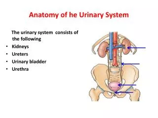
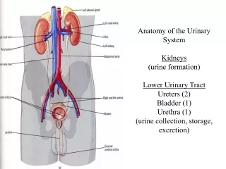
The Urinary System • Kidneys, ureters, urinary bladder & urethra • Urine flows from each kidney, down its ureter to the bladder and to the outside via the urethra • Filter the blood and return most of water and solutes to the bloodstream and excrete the waste products. Solution 溶解
Kidney Location The kidneys lie on the posterior abdominal all on either side of the vertebral column. The distance between the inferior extremities is wider than that between the superior extremities. The right kidney is lower than the left one because of the large size of the lobe of the live. The superior extremity of the left kidney is at the level of the inferior border of the body of the 11th thoracic vertebra, the inferior extremity of the left kidney is at the level with the inferior border of the body of the 2nd lumbar vertebra.
The superior extremity of the right kidney is at the level with the superior border of the body of the 12th thoracic vertebra, and the inferior extremity is at the level with the superior border of the body of the third lumbar vertebra. The renal hilum is at the level of the first lumbar vertebra, 5cm lateral to the midline of the body. The area between the 12th rib and the lateral border of the erector spinae is called the renal region in clinic. The location of The kidneys varies in different cases. General in female it is lower than in man. Location of the kidney
External Anatomy of Kidney 2 surfaces anterior: convex posterior: plane 2 borders medial: renal hilum lateral: convex renal sinus and renal pedicle 2 poles : superior: broad & thin inferior: narrow & thick
The hilum transmits the renal vessels, nerves and the part of the pelvis. The renal hilum leads into a central recess named the renal sinus. The renal sinus is filled with the branches of the renal artery and vein, nerves, lymphatic vessels, minor renal calices, major renal calices, renal pelvis and adipose tissue. Renal pedicle The structures which pass through the renal hilum are enclosed together by connective tissue.
Internal structures Renal columns Cortex It is composed of the renal glomeruli and renal tubules Medulla Renal pyramids(base and apices) 2 or 3 (7 to 12 in each kideney)Renal papilla (Papillary foramina10to 30 in each papillae) 2 or 3 Minor renal calices Renal pelvis Major renal calices
Coverings Fibrous capsule Fatty renal capsule Renal fascia Adipose capsule Renal fascia Anterior layer Fibrous capsule Posterior layer
Blood vessels & ureter enter hilum of kidney • Renal capsule : from outer to inner is the renal fascia, adipose capsule and the fibrous capsule. • Adipose capsule that helps protect from trauma • Renal fascia = dense, irregular connective tissue that holds against back body wall
Internal Anatomy of Kidney • What is the difference between renal hilum & renal sinus? • Outline a major calyx & the border between cortex & medulla.
Renal segments • ①Superior segment • ②Superior anterior segment③inferior segment • ④Inferior anterior segment⑤Posterior segment Renal segments Cast of kidney
Blood & Nerve Supply of Kidney • Abundantly supplied with blood vessels • receive 25% of resting cardiac output via renal arteries • Functions of different capillary beds • glomerular capillaries where filtration of blood occurs • vasoconstriction & vasodilation of afferent & efferent arterioles produce large changes in renal filtration • peritubular capillaries that carry away reabsorbed substances from filtration. • vasa recta supplies nutrients to medulla without disrupting its osmolarity form • Sympathetic vasomotor nerves regulate blood flow & renal resistance by altering arterioles Glomerulus 球 osmolarity 渗透性
Overview of Kidney Functions • Regulation of blood ionic composition • Na+, K+, Ca+2, Cl- and phosphate ions • Regulation of blood pH, osmolarity & glucose • Regulation of blood volume • conserving or eliminating water • Regulation of blood pressure • secreting the enzyme renin ( 血管紧张肽) • adjusting renal resistance • Release of erythropoietin(红细胞生成素)& calcitriol(降钙素) • Excretion of wastes & foreign substances • Na+ sodium ions K+ potassium ions Ca+2 calcium ions Cl- chlorine ions
The renal anomalies • ①The early pelvic position of the kidneys may persists. • ②the kidneys are connected by a transverse bridge of renal tissue forming the “horseshoe kidney”. • ③the kidney of one side may be congenital absence or imperfect development. • ④teo kidneys may be occur on the same side. • ⑤single or multiple congenital renal cysts may also be present.
Renal cysts horseshoe Variation Pelvic position
Diuretics • Substances that slow renal reabsorption of water & cause diuresis (increased urine flow rate) • caffeine which inhibits Na+ reabsorption • alcohol which inhibits secretion of ADH) • Diuresis (利尿) Na+ sodium ions • Caffeine (咖啡因) ADH antidiuretic hormone
Ureters • It is muscular tube, about 20 to 30 cm in length. • Varies in diameter from 1-10 mm. • Extends from renal pelvis to bladder. • Retroperitoneal • Enters posterior wall of bladder ( intramural seg.) • Physiological valve only • bladder wall compresses arterial opening as it expands during filling • flow results from peristalsis, gravity & hydrostatic pressure Trigone
Ureters • Three parts • The abdominal part • It is continuous with the pelvis superiorly, at the superior pelvic aperture, the left ureter crosses through the terminal part of the left common iliac artery anteriorly, and the right ureter passes the beginning of the right external iliac artery anteriorly, to continue with the pelvic part.
Ureters • The pelvic part • It passes downwords along the lateral wall of the lesser pelvis, then turns medially at the level of ischial spine to the base of the urinary bladder. At here the ductus deferens crosses it anteriorly to its medial sode in the male. In the femal the pelvic part of the ureter turns downwords, forwords and medially. At the level of ischial spine, 2cm lateral to the cervix of uterus, it is crosses anteriorly by the uterine vessels. It then runs along the lateral side and the anterior surface of the vagina to the base of the urinary bladder.
Ureters • The intramural part • It passes obliquely through the wall of the urinary bladder for 1.5cm long, before opening into the superolateral angle of fundus of bladder. Where the bladder is filled up, the intramural part seces as a valve to prevent the backflow of urine.
Ureters • Three constrited parts • The ureters are not uniform in calibre. Three constricted parts are the places where the renal stones stay usually. • ①at the junction of the ureter and the renal pelvis. • ②at the point where ureter crosses the iliac vessels. • ③intramural part. • These constricted areas are potential sites of obstruction by ureteric stones.
Urinary Bladder • Hollow, distensible muscular organ with capacity of 700 - 800 milliliter. • Trigone is smooth flat area bordered by 2 ureteral openings and one urethral opening
Apex Body Fundus Neck External features
Location of urinary bladder In the adult the empty bladder is entirely within the lesser pelvis, but as it becomes distended it upwards and forwards into the abdominal cavity. At the baby, the empty bladder is spindle-shaped , and lies at a relatively higher level than in the adult. It is a interperitoneal organ, only the superior surface and upper portion of the inferolateral surface are covered by peritoneum, which is reflected from the lateral wall of the pelvis and from the anterior abdominal wall just above the level of the pubic symphysis when the bladder is empty.
As the bladder is filled by urine, the superior surface of the bladder enlarges and bulges upward into the abdominal cavity, the peritoneal covering is peeled off away from the lower part of the anterior abdominal wall, and reflection of the peritoneum becomes higher. So that the bladder contracts directly with the anterior abdominal wall. Therefore the puncture of bladder can be performed just above the pubic symphysis within injuring the peritoneum.
The urethra • The urethra extends from internal urethral orifice of the urinary bladder to the external urethral orifice. It is very different in male and femal. • The male urethra serves a common tube for urinary and genital systems. It may be divided into three parts. It will be described in the genital system in detail. • The femal urethra
The femal uretha • It is membranous canal, about 5cm long, extending from the bladder to external urethral orifice in the vaginal vestibule. Its diameter is about 6mm. It is dorsal to the pubic symphysis and ventral to the anterior wall of the vagina. Its direction is obliquely and forward and slightly curved. It perorates the pelvic floor and uroginital diaphragm, and its external orifice is situated just anterior to the vaginal orifice and about 2.5cm posteroinferior to the glans of clitoris. Where the urethra passes through the uroginital diaphragm, the striated muscle of uroginital diaphragm forms the urethrovaginal sphincter. Many small urethral glands open into the urethra. The largest of these is the paraurethral glands, the duct of which open into the vaginal vsetibule on either side of the external orifice of urethra.
The renal transplantation • The kidney can be removed from the donor without damaging the suprarenal gland because of the weak septum of renal fascia that separates the kidney from this gland. Renal transplantation is now an established operation for the treatment of selected cases of chronic renal failure. The site artery and vein are joined to the external iliac artery and vein, respectively, and the ureter is sutured into the urinary bladder.