Blood Pressure
An Individuals blood pressure is a standard clinical measurement Is considered a good indicator of the status of the cardiovascular system.

Blood Pressure
E N D
Presentation Transcript
An Individuals blood pressure is a standard clinical measurement • Is considered a good indicator of the status of the cardiovascular system. • Blood pressure values in the various chambers of the heart and in the peripheral vascular system help the physician determine the functional integrity of the cardio vascular system. Blood Pressure
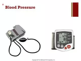
Direct (invasive) • Extravascular Method • The vascular pressure is coupled to an external sensor element via a liquid filled catheter. • [Catheter – is a long tube introduced into the heart or a major vessel by way of a superficial vein or artery.] • 2. Intravascular • A sensor is placed into the tip of a catheter that is placed in the vascular system. • Indirect (non invasive) • Sphygmomanometer • Consists of an inflatable pressure cuff and a manometer to measure the pressure in the cuff. Blood Pressure Measurement
The extra vascular sensor system is made up of a catheter. • The catheter is connected to a three way stopcock and then to a pressure sensor • It is filled with a saline-heparin solution. • It must be flushed with solution every few minutes to prevent blood clotting at the tip. Direct MeasurementExtra Vascular
Physician inserts the catheter • Either by means of a surgical cut-down, which exposes the artery or vein. • or by means of percutaneous insertion which involves the use of a special needle or guide-wire technique. • Blood pressure is transmitted via the catheter column to the sensor and finally to the diaphragm which is deflected. • The displacement of the diaphragm is sensed electronically. Direct MeasurementExtra Vascular contd…
Direct MeasurementExtra Vascular contd… • Disadvantages • The frequency response of the catheter-sensor system is limited by the hydraulic properties of the system. • Creates time delay in detection of pressures when a pressure pulse is transmitted.
Direct MeasurementIntravascular • The sensor is placed at the tip of the catheter. • Enables the physician to obtain a high frequency response in detection of pressures at the tip of the catheter. • Types of sensors • Strain-gage systems • bonded onto a flexible diaphragm at the catheter tip. • Fibre-optic device • Measures the displacement of the diaphragm optically by varying reflection of light from the back of the deflecting diaphragm.
Consists of strain-sensitive gages which are firmly bonded with an adhesive to the membrane or diaphragm whose movement is to be recorded. • Made by taking a length of a very thin wire or foil which is formed into a grid pattern and bonded to a backing material. • Is then attached to the diaphragm. • Deflection of the diaphragm causes corresponding strain in the wire gage. • Causes a corresponding change in the resistance which is proportional to the pressure. Bonded Strain Gage pressure transducer
Measures the displacement of the diaphragm optically by the varying reflection of light from the back of the deflecting diaphragm. • Inherently safer electrically Fiber optic type pressure transducer
Using Fourier Analysis techniques in the quantification of pressure and flow. • Blood pressure pulse can be divided into its fundamental component (of the same frequency as the blood pressure wave) and its significant harmonics. • Analysis of the frequency components of the pulse yield more information on arterial properties. Harmonic Analysis of Blood Pressure Waveforms
Testing technique for measuring the transient response of the catheter-sensor system
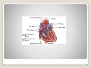
For determining the function of • Capillary bed • Right side of the heart • The central venous pressure is measured in the central vein or in the right atrium. • It fluctuates above and below atmospheric pressure as the subject breathes. • The reference level for venous pressure is at the right atrium. • Central venous pressure is an important indicator of myocardial performance Venous pressure
Monitored for assessing proper therapy for • heart dysfunction • Shock • Hypovolemic(Of or relating to a decrease in the volume of circulating blood)or hypervolemic States • Circulatory failure Central venous pressure
Physicians usually measure steady state or mean venous pressure by making a percutaneous venous puncture with a large bore needle, inserting a catheter through the needle . • Needle is then removed.
Continuous dynamic measurements is made by connecting a high sensitive pressure sensor to the venous catheter. • Normal venous pressure values range widely from 0 to 1.2 kPa with a mean pressure of 0.5 kPa(0 – 12cm H2O). Central venous pressure
Heart sounds are vibrations or sounds due to the acceleration or deceleration of blood. • Murmurs are vibrations or sounds due to blood turbulence. • The technique of listening to sounds produced by the organs and vessels of the body is called auscultation. Heart Sounds
With each heartbeat, the normal heart produces two distinct sounds that are audible in the stethoscope – often described as “lub-dub” • The “lub” is caused by the closure of atrioventricular valves and is called the first heart sound • occurs approximately at the time of QRS complex of the ECG and just before ventricular systole. • The “dub” part of the heart sounds is called the second heart sound and is caused by the closing of the semilunar valves • Which closes at the end of the systole, just before the atrioventricular valve opens. • Occurs at the time of the end of the T wave of the ECG • The third heart sound attributed to the sudden termination of the rapid filling phase of the ventricles from the atria and the associated vibration of the ventricular muscle walls., which are relaxed. Heart Sounds …
Fourth or atrial heart sound – not audible , can be recorded by phonocardiogram, due to atria contract
There are optimal recording sites for the various heart sounds. Auscultation Techniques
Heart sounds and murmurs have extremely small amplitudes with frequencies from 0.1 to 2000 Hz. • Thus the recording device must be carefully selected for wide band frequency response characteristics. • Specially designed acoustically quiet environment is needed for noise free recording of heart sounds. Auscultation Techniques…
Mechanical stethoscopes amplifies sound because of Standing wave phenomenon. • Firm application of the chest piece makes the diaphragm taut with pressure thereby causing an attenuation of low frequencies. • Loose-fitting earpiece cause leakage which reduces the coupling between the chest wall and the ear. • Electronics stethoscopes has selectable frequency response characteristics ranging from “ideal” flat-response to selectable band-pass response. Stethoscope
A Phonocardiogram is a recording of the heart sounds and murmurs. • Eliminates subjective interpretation of the heart sounds • Enables evaluation of the heart sounds and murmurs with respect to the electric and mechanical events in the cardiac cycle. • Evaluation of the result is based on the basis of changes in the wave shape and various timing parameters. Phonocardiogram
The process of introducing a catheter into the heart for diagnosis. • Used to asses hemodynamic (circulation of the blood and the forces involved) function and cardiovascular structure. • Performed during most of the heart surgeries. • Performed in specialized laboratories outfitted with x-ray equipment for visualizing heart structures and the position of various pressure catheters. Cardiac Catheterization
A radiopaque die is injected into the ventricles or aorta through the catheter for assessing the ventricular or aortic function • Pressures in all four chambers of the heart and in the great vessels can be measured by positioning the catheters in such a way to recognize the characteristics pressure waveforms. Cardiac Catheterization …
Angiographic visualization is an essential tool used to evaluate cardiac structure. • Specially designed catheters and power injectors are used in order that a bolus of contrast material can be delivered rapidly into the appropriate vessel or heart chamber. • During catheterization cardiac catheterization frequently occur. Clinics must have a functional defbrillaltor Angiography
Angeography Types • Left & Right ventricle – ventriculography • Coronary arteries – coronoryangeography • Pulmonary artery – pulmonary angeography • Aorta- aortography
Surgical procedure to repair a damaged blood vessel or unblock a coronary artery. • PTCA – PercuntaneousTransluenal Coronary Angeoplasty Angioplasty