Chest Trauma
Chest Trauma . Surgery department № 2 DSMA. Introduction. Chest trauma is often sudden and dramatic Accounts for 25% of all trauma deaths 2/3 of deaths occur after reaching hospital

Chest Trauma
E N D
Presentation Transcript
Chest Trauma Surgery department № 2 DSMA
Introduction Chest trauma is often sudden and dramatic Accounts for 25% of all trauma deaths 2/3 of deaths occur after reaching hospital Serious pathological consequnces: -hypoxia, hypovolaemia, myocardial failure
Mechanism of Injury Penetrating injuries E.g. stab wounds etc. Primarily peripheral lung Haemothorax Pneumothorax Cardiac, great vessel or oesophageal injury
Blunt injuries Either: direct blow (e.g. rib fracture) deceleration injury compression injury Rib fracture is the most common sign of blunt thoracic trauma Fracture of scapula, sternum, or first rib suggests massive force of injury
Chest wall injuries Rib fractures Flail chest Open pneumothorax
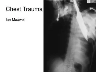
Rib fractures Most common thoracic injury Localised pain, tenderness, crepitus CXR to exclude other injuries Analgesia avoid taping Underestimation of effect Upper ribs, clavicle or scapula fracture: suspect vascular injury
Flail chest Multiple rib fractures produce a mobile fragment which moves paradoxically with respiration Significant force required Usually diagnosed clinically Rx: ABC Analgesia
Open pneumothorax Defect in chest wall provides a direct communication between the pleural space and the environment Lung collapse and paroxysmal shifting of mediastinum with each respiratory effort ± tension pneumothorax “Sucking chest wound” Rx: ABCs…closure of wound…chest drain
Lung injury Pulmonary contusion Pneumothorax Haemothorax Parenchymal injury Trachea and bronchial injuries Pneumomediastinum
Pneumothorax Air in the pleural cavity Blunt or penetrating injury that disrupts the parietal or visceral pleura Unilateral signs: movement and breath sounds, resonant to percussion Confirmed by CXR Rx: chest drain
Pneumothorax classification By side: • left or right • in both side By lung collapse degree: • Partial (paracostal) • Subtotal (smaller than 2/3 of lung volume) • Total (more than 2/3 of lung volume) By mechanism of formation: - open - closed - tension
Tension pneumothorax Air enters pleural space and cannot escape P/C: chest pain, dyspnoea Dx: - respiratory distress - tracheal deviation (away) - absence of breath sounds - distended neck veins - hypotension
Surgical emergency Rx: emergency decompression before CXR Either large bore cannula in 2nd ICS, MCL or insert chest tube CXR to confirm site of insertion
Haemothorax Blunt or penetrating trauma Requires rapid decompression and fluid resuscitation May require surgical intervention Clinically: hypovolaemia absence of breath sounds dullness to percussion CXR may be confused with collapse Decompression always by chest catchment in 7 ICS on middle or posterior axillary line
Hemothorax classification By side: • left or right • in both side By blood lost volume : • Small (< 10% of BCV, or <500 ml) • Middle (10-20 % of BCV, or 500-1000ml) • Big (10-20 % of BCV, or 500-1000ml) • Total ( > 40 % of BCV, or >2000ml) By bleeding presence: - stopped (Reviloi – Gregoire test negative) - continues (Reviloi – Gregoire test positive) By clots presence: - clotted - unclotted By infection complication presence: - non-infected - infected
Indication for urgent thoracotomy • In pneumothorax: Absence of active air catchment during more than 2 days (presence of pneumothoraz on CXR) • In hemothorax: Evacuation of > 1000ml blood simultaneously or bleeding continues during 4 hours with blood loss > 200 ml per hour
Heart, Aorta & Diaphragm Blunt cardiac injury - contusion - ventricular, septal or valvular rupture Cardiac tamponade Ruptured thoracic aorta Diaphragmatic rupture
Cardiac Tamponade Blood in the pericardial sac Most frequently penetrating injuries Shock, JVP, PEA, pulsus paradoxus Classically, Beck’s triad: - distended neck veins - muffled heart sounds - hypotension Rx: Volume resuscitation Pericardiocentesis
Aortic rupture Usually blunt trauma involving deceleration forces; especially RTAs ~90% die within minutes Most common site near ligamentum arteriosum Dx: clinical suspicion, CXR, aortography, contrast CT or TOE Rx: surgical…poor prognosis
Iatrogenic trauma NG tubes: - coiling - endobronchial placement - pneumothorax Chest tubes: - subcutaneous - intraparenchymal - intrafissural Central lines: - neck - coronary sinus - pneumothorax
Chest trauma: summary Common Serious Primary goal is to provide oxygen to vital organs Remember Airway Breathing Circulation Be alert to change in clinical condition