Download
1 / 28
290 likes | 536 Views
Commitment to complete T cell activation requires many hours: MHC-peptide ligands which dissociate early can lead to partial activation. No activation. Partial activation. time. Complete T cell activation. Figure 8-10.

E N D
Commitment to complete T cell activation requires many hours: MHC-peptide ligands which dissociate early can lead to partial activation No activation Partial activation time Complete T cell activation
Figure 8-10 Co-stimulation provides a second signal which is necessary for activation of naïve lymphocytes
Figure 9-5 B cell activation is regulated by co-stimulatory signals provided by CD40. The ligand for CD40 (CD40L) is induced on activated T cells.
CD40 stimulation also further activates APCs. This stimulates APC function, including greater B7 expression. High levels of co-stimulatory activity promotes the activation of CD8+ T cells which are strongly dependent on co-stimulation. CD4+ T cell CD8+ T cell CD40L B7 B7 B7 B7 CD40 B7 APC APC B7
Figure 11-12 Hyper IgM syndrome is commonly due to a defective allele of the CD40 ligand gene Characteristics: 1. Relatively high levels of IgM and the absence of germinal centers. Increased susceptibility to p.carinii infection 3. Reduced T cell response
Stimulation in the absence of a co-stimulatory signal inactivates the T cell, while a co-stimulatory signal alone has no effect
Once anergized, T cells fail to response to signals which would normally lead to activation. This is a means of inducing tolerance to self antigens.
Stimulation with a partial agonist, or disruption of the CD4 - MHCII interaction, can lead to anergy in some cases. anergizing activating
TCR Y Y Y Y CD28 IL-2R IL-2 Activated Induction of anergy depends upon a signal from the TCR and can be blocked by inhibitors of the phosphatidylinositol pathway TCR TCR Cyclosporin A Y Y IL-2R Anergized No response
In vitro restimulation assay i.v. inject 1µg p33 p33 + 3 days 2 days TCR Tg a LCMV p33 3H-thymidine incorporation + CD28+/+ or CD28-/- spleen 1000 APC 100 Proliferation (% of response from uninjected animals) 10 1 CD28+ CD28- CD28+ CD28- no IL-2 + IL-2 Lack of CD28 promotes anergy induction in vivo which can be reversed by IL-2 +/- IL-2 adapted from Bachmann, et al 1997
CD28 provides 2nd signal The B7 ligands (CD80/CD86) on APCs provide activation signals to the naïve T cell that are required for the production of IL-2 and prevent the induction of anergy
Figure 8-18 part 2 of 2 The APCs have specialized functions, but the co-stimulatory activity of all three types is regulated
Figure 8-15 Dendritic cells first encounter antigen in peripheral tissues, and then undergo maturation while migrating to draining lymph nodes. Increased expression of B7 on mature dendritic cells makes them excellent APCs for naïve T cells.
Figure 8-16 Activation of APCs by microbial products or cytokines induces expression of B7. Co-stimulation requires a “danger” signal.
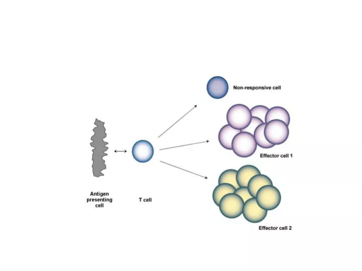
Lymphocyte activation is a self-limited process death memory
Figure 8-12 Induction of CTLA-4 limits the T cell response by blocking the co-stimulatory function of CD28. Mice with a CTLA-4 deletion exhibit massive lymphoproliferation and lethality.
Some cell surface receptors have Immunoreceptor Tyrosine-based Inhibitory Motifs (ITIMs) within their cytoplasmic domain which deliver a negative signal
Phosphatases containing SH2 domains, SHP-1 and SHIP-1, can bind phosphorylated ITIMs and are involved in inhibiting immunoreceptor signaling
Co-aggregation of the BCR and FcRIIB receptors by antibody saturated antigen blocks activation. BCR associated kinases phosphorylate FcR ITIMs allowing recruitment of the SHIP 5’ inositol phosphatase. SHIP activity reduces PIP3 levels reducing binding of PH domain proteins and activation of signaling pathways.
Figure 6-24 Induction of the Fas ligand on activated T cells limits T cell expansion through Fas mediated apoptosis. This is known as activation induced cell death.
Autoimmune Lymphoproliferative Syndrome (ALPS) • Characterized by lymphadenopathy and splenomegaly with greatly increased numbers of lymphocytes. • Associated with autoimmune cytopenia • most patients have a dominant mutation effecting the Fas gene
Your research advisor has identified a line of mice which are immunodeficent due to a defective T cell response. You have been asked to determine the basis for this defect. a). Examining lymph nodes using a new technique, intravital imaging, you are able to detect T lymphocytes rolling on the surface of high endothelial venules, but very few lymphocytes are found inside the nodes. What protein, if defective, could account for this observation? b). Analysis of different cell types shows that the defect responsible for the immunodeficiency specifically effects T cells. For further analysis you generate a TCR transgenic mouse line which has the immunodeficiency. Mixing together naive T cells, the antigen, and APCs, you observe only a few T cell – APC pairs under the microscope. In contrast, many T cell – APC pairs are observed using naïve TCR transgenic T cells from non-immunodeficient animals. If you fail to include antigen, no pairs are seen. You suspect defects in adhesion molecules, but your advisor says that purified adhesion molecule ligands bind equally well to naïve T cells from normal and immunodeficient animals. How do you explain the defect in interaction between T cells and APCs? c). Draw a simple picture indicating the relative position of the TCR and adhesion molecules at the interface between an APC and a responding T cell once the TCR is stably engaged.
The Centers for Disease Control has identified a new T lymphotrophic virus. Eager to help combat an epidemic, you start doing research to figure out how it effects the T cells it infects. a). You determine that this virus encodes a membrane raft-associated protein which promotes viral replication by directly binding and activating the ZAP-70 tyrosine kinase. Do you expect this protein to effect ITAM phosphorylation? NFAT activation? Akt activation? For each case, explain your answer. b). Herpesvirus saimiri strain 484C encodes a protein which activates the Src kinase Lck. How would you expect this protein to effect ITAM phosphorylation? NFAT activation? Akt activation?Explain why, or why not c). Like the HIV virus, the new T lymphotrophic virus enters the cell in part by binding to CD4. You propose generating a reagent which blocks viral entry by binding to the extracellular domain of CD4. Your antiviral agent proves to be an excellent inhibitor of viral infection! However, it also appears to be an excellent immunosupressive agent, capable of blocking T cell activation. Why is activation blocked?
One mechanism used by the immune system to avoid reactivity to self antigens is the induction of anergy in naïve T cells. Describe conditions where naïve T cells are anergized rather than activated: what molecule on the T cell, when stimulated, promotes activation, while a failure to stimulate it may lead to anergy? What is produced as a result of this stimulation? (Do not discuss signaling mechanisms). What molecules on antigen presenting cells help determine whether stimulation of naïve T cells leads to activation or anergy? What determines the expression of these molecules on APCs? If an APC was unable to provide a co-stimulatory signal, and did not present a peptide recognized by naïve T cells, would the T cells be anergized following interaction with the APC?
You are attempting to stimulate T cells from a TCR transgenic mouse with a partial agonist peptide and APCs. You find that there is little T cell proliferation. a). Should you attempt to increase the response by adding anti-CD28 antibodies to improve the costimulatory signal, or treat the cells with a reagent to increase the activity of adhesion molecules? b). When you use the normal agonist you are able to identify a new cell-surface protein which is induced by T cell activation and co-localizes with CD28. This protein associates with the SHIP 5’ inositol phosphatase. What effect will this have on CD28 signaling pathways? Explain your answer. If this protein co-localized with CD40 in B cells, what effect would it have on CD40 co-stimulation?