Download
1 / 53
540 likes | 690 Views
THYROID GLAND, PARATHYROID GLAND, ADRENAL GLAND, PINEAL GLAND, PANCREAS Dr. Anna L. Kiss Semmelweis University Department of Anatomy, Histology and Embryology 2018. ENDOCRINE GLANDS. Pineal gland. Hypophysis. Parathyroid gland. Thyroid gland. Adrenal gland. Endocrine pancreas. Gonads.

E N D
THYROID GLAND, PARATHYROID GLAND, ADRENAL GLAND, PINEAL GLAND, PANCREAS Dr. Anna L. Kiss Semmelweis University Department of Anatomy, Histology and Embryology 2018
ENDOCRINE GLANDS Pineal gland Hypophysis Parathyroid gland Thyroid gland Adrenal gland Endocrine pancreas Gonads
ENDOKRIN MIRIGYEK 1.) Directed by the hypothalamo-hypophyseal system: thyroid gland adrenal cortex gonads 2.) NOT directed by the hypothalamo-hypophyseal system: parathyroid adrenal medulla Langerhans islands corpus pineale
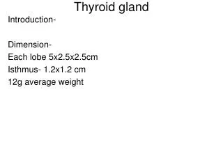
THYROID GLAND anatomy Larynx Lobes Isthmus Trachea
THYROID GLAND anatomy It develops behind the buccopharyngeal membrane from the thyroglossal duct + neural crest cells Pyramidal lobe (~40%) Lobes Isthmus
THYROID GLAND histology Capsule: outer loose connective tissue inner fibroelastic tissue Connective tissue septa: irregular lobules Parenchyma: follicles: spheres (0.2-1 mm in diameter) simple epithelium on basal lamina: follicular cells central colloid core parafollicular cells (C cells) Stroma: loose connective tissue with: dense plexus of fenestrated capillaries lymphatic vessels symphathetic nerve fibers
THYROID GLAND histology Follicular cells: from low cuboidal to columnar depending on the functional state round nucleus, prominent nucleolus basophilic cytoplasm: basal rER supranuclear Golgi apparatus apical small secretory vesicles apical endosomes: colloidal resorption droplets lysosomes apical microvilli Colloid: thyroglobulin: - is an iodinated glycoprotein (660 kDa) ~120 tyrosine residues inactively stored thyroid hormones Thyroid hormones: iodinated amino acid derivatives (tyrosine)
THYROID GLAND histology Thyroid hormone synthesis: 1. synthesis of thyroglobulin and thyroperoxidase in rER, modification in Golgi, secretion on the apical plasma membrane 2. uptake of iodide from blood on the basal plasma membrane (Na+/J- symporter); secretion on the apical membrane (J-/Cl- transporter) oxydation of iodide to iodine (thyroperoxidase!) in the colloid 3. iodination of thyroglobulin: monoiodotyrosine (MIT) and diiodotyrosine (DIT) formation 4. triiodothyronine (T3) and thyroxine (T4) formation 5. resorption of colloid; vesicles fused with lysosomes 6. release of T3 and T4 into blood
THYROID GLAND histology Septum Capsule
THYROID GLAND histology Microvilli Follicular cell Colloid Site of thyroglobulin resorption
THYROID GLAND function Role of thyroid hormones: critical for normal growth and development increase of basal metabolic rate Regulation of thyroid hormone secretion: by hypothalamo-hypophyseal TRH-TSH system Hypothyreodism: infancy, early childhood: impaired growth and CNS development – Cretenism adult: slowing of physical and mental activity – Myxedema
Hypothyreosis: infancy, early childhood: - dwarfism - cretenism adult: physical and mental slowing down goiter (hypofunctional: connective tissue!) myxoedema
THYROID GLAND function Hyperthyreodism: Basedow disease hypermetabolic state exoftalmus goiter
THYROID GLAND-PARAFOLLICULAR CELLS Parafollicularcells (C cells): located: • in theperiphery of follicles: withinthebasallamina neverreachthe lumen • in clusters in theinterfollicularstroma pale-stainingcytoplasm(C forclear): prominentGolgi apparatus smallsecretoryvesicles secretecalcitonin: 32-amino acidpolypeptide
THYROID GLAND-PARAFOLLICULAR CELLS histology Parafollicular cell Follicular cell Colloid
THYROID GLAND-PARAFOLLICULAR CELLS histology Parafollicular cells
THYROID GLAND-PARAFOLLICULAR CELLS function Role of calcitonin: decreases blood calcium level: ↓ Ca2+ resorption from the bone ↑ calcium deposition into bones Regulation of calcitonin secretion: by blood calcium level hypophysis-independent No clinical symptoms
PARATHYROID GLAND anatomy Pharynx Thyroid gland Parathyroid glands Oesophagus
PARATHYROID GLAND anatomy Shape: ovoid or lenticular Colour: yellowish-brown Position: behind the posterior lobar border of thyroid gland, within the capsule Dimensions: length: 3-6 mm width: 2-4 mm thickness: 1-2 mm Weight: 0.1-0.2 g
PARATHYROID GLAND histology Capsule: thin connective tissue Connective tissue septa: poorly defined lobules Parenchyma: cords of epithelial cells: principal (chief) cells oxyphil cells Stroma: loose connective tissue with: dense plexus of fenestrated capillaries lymphatic vessels adipose tissue increases till adulthood (up to 30%)
PARATHYROID GLAND histology Principal (chief cells): predominant cell type small polygonal cells large spherical nucleus pale, slightly eosinophilic cytoplasm (activity dependent): Golgi apparatus in active cells small secretory vesicles in active cells glycogen in inactive cells lipid droplets lipofuscin granules secrete parathormone (PTH): 84-amino acidpolypeptide
PARATHYROID GLAND histology Oxyphil cells: minor cell population, singly or in clusters large rounded cells smaller darker nucleus acidophilic cytoplasm: large number of mitochondria no secretory vesicles little rER produce?
PARATHYROID GLAND histology Septum Adipose tissue Capsule
PARATHYROID GLAND histology Oxyphil cell Principal cell (inactive) Principal cell (active)
PARATHYROID GLAND function Role of parathormone (PTH): increases blood calcium level: ↑ bone resorption ↑ calcium reabsorption in kidneys ↑ formation of active vitamine D3 ↑ intestinal absorption of calcium decreases blood phosphate level: ↑ phosphate excretion in kidneys Regulation of parathormone secretion: by blood calcium level hypophysis-independent
PARATHYROID GLAND function Hypoparathyroidism: hypocalcemia → tetany Hyperparathyroidism: hypercalcemia, osteoporosis
ADRENAL GLAND anatomy Right adrenal gland Left adrenal gland
ADRENAL GLAND anatomy Shape: right: pyramidal left: semilunar Colour: golden yellow Position: superior and slightly anterior to the kidneys left adrenal gland is more inferior
ADRENAL GLAND histology Capsule: thick, dense irregular connective tissue Delicate connective tissue trabecules Parenchyma: cortex: constitutes 90% of adrenal weight yellowish in colour secretion of steroid hormones medulla: constitutes 10% of adrenal weight dark red or brownish in colour secretion of catecholamines Stroma: loose connective tissue with: dense plexus of fenestrated capillaries preganglionic sympathetic fibers in medulla
ADRENAL CORTEX histology Zonation: zona glomerulosa: narrow outer zone 15% of cortical volume closely packed ovoid or round clusters of: small columnar or pyramidal cells zona fasciculata: thick middle zone 80% of cortical volume long straight cords (1-2 cell thick) of: large polyhedral cells zona reticularis: thin inner zone 5% of cortical volume anastomosing cords of: smaller cells
ADRENAL GLAND histology Capsule Zona glomerulosa Zona fasciculata Zona reticularis Medulla
ADRENAL CORTEX histology Zona glomerulosa cells: small columnar or pyramidal cells densely stained spherical nucleus pale cytoplasm: abundant sER numerous large mitochondria multiple Golgi complexes free ribosomes sparse lipid droplets secrete mineralocorticoids (aldosterone)
ADRENAL CORTEX histology Zona fasciculata cells: large polyhedral cells (binucleate) lightly stained spherical nucleus pale-staining vacuolated cytoplasm: abundant sER numerous tubular mitochondria well-developed Golgi apparatus rER numerous lipid droplets secrete glucocorticoids (cortisol)
ADRENAL CORTEX histology Zona reticularis cells: smaller cells darkly stained spherical nucleus darker cytoplasm: large lipofuscin granules well-developed sER numerous tubularmitochondria few lipid droplets secrete weak androgens (DHEA)
ADRENAL GLAND histology Capsule Zona glomerulosa Zona fasciculata
ADRENAL GLAND histology Zona fasciculata cell Sinusoid
ADRENAL MEDULLA histology Structure: ovoid clusters and interconnecting cords of: large columnar cells: chromaffin cells ganglion cells
ADRENAL MEDULLA histology Chromaffin cells: innervated by preganglionic sympathetic fibers!!! large columnar (epitheloid) cells lightly stained large nucleus basophilic cytoplasm: rER Golgi apparatus secretory vesicles: dense core vesicles (large): norepinephrine small secretory vesicles: epinephrine Ganglion cells: characteristic neurons (innervated by preganglionic sympathetic fibers)
ADRENAL GLAND histology Zona reticularis Medulla
ADRENAL GLAND histology Ganglion cell Chromaffin cell
ADRENAL GLAND blood supply Components: capsular arteries ↓ ↓ cortical arterioles medullary arterioles ↓ ↓ cortical sinusoids ↓ medullary sinusoids ↓ medullary veins Medulla receives blood from: medullary arterioles cortical sinusoids t. media: longitudinally oriented bundles of smooth muscle cells adrenomedullary veins
ADRENAL CORTEX function Role of mineralocorticoids: control of electrolyte homeostasis and water balance: ↑ sodium reabsorption in kidneys ↑ potassium excretion in kidneys Regulation of mineralocorticoid secretion: by renin-angiotensin system Adrenocortical insufficiency: hyperkalemia, hyponatremia, hypovolemia: hypotension leading to shock Hyperaldosteronism: hypokalemia, hypertension
ADRENAL CORTEX function Role of glucocorticoids: increase glucose availability: ↑ gluconeogenesis ↑ glycogenesis ↑ protein and lipid degradation suppress inflammatory response Regulation of glucocorticoid secretion: by hypthalamo-hypophyseal CRH-ACTH system Hypercortisolism: hyperglycemia, increased protein degradation, hypertension, cental adipose deposition, increased risk of infections
ADRENAL MEDULLA function Role of catecholamines: prepare the body for „fight-or-flight” response: ↑ heart rate ↑ blood pressure ↑ rate of respiration ↑ sweating ↓ digestion Regulation of catecholamine secretion: by sympathetic nervous system conversion of norepinephrine to epinephrine is induced by cortical glucocorticoids Catecholamine overproduction: hypertension
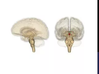
PINEAL GLAND anatomy Pineal gland
PINEAL GLAND histology Pia mater covering Connective tissue septa: lobules Glandular tissue: clumps or cords of: pinealocytes interstitial (glial) cells: similar to astrocytes neurons: processes to habenular nucleus (medial), pretectal nuclei, hypothalamus Fenestrated capillaries Brain sand (corpora arenacea): concentric basophilic concretions: calcification on secreted proteins increase in number with age reference point on radiographic and CT images
PINEAL GLAND histology Pia mater Pineal recess Brain sand Posterior commissure
PINEAL GLAND histology Pinealocytes: predominant cell type cells with cytoplasmic processes deeply infolded large nucleus, 1-2 nucleolus pale cytoplasm: few organelles dense core secretory vesicles in cytoplasmic processes processes are associated with: capillaries – neuroendocrine activity pineal neurons: axo-dendritic synapses secrete melatonin: indolamine
PINEAL GLAND histology Pinealocytes Glial cell
PINEAL GLAND histology Neuron