Download
1 / 1
10 likes | 203 Views
Strategies For Generating Microarray Data From LCM Derived Breast Tissue RNA S. Tighe, T. Casey, L. Lintault, J.White, J. Eneman, T. Hunter, M. A. Chaudhry, K. Plaut, and H Muss. Vermont Cancer Center and University of Vermont Burlington, Vermont . Abstract. Results.

E N D
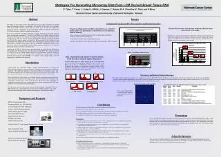
Strategies For Generating Microarray Data From LCM Derived Breast Tissue RNA S. Tighe, T. Casey, L. Lintault, J.White, J. Eneman, T. Hunter, M. A. Chaudhry, K. Plaut, and H Muss. Vermont Cancer Center and University of Vermont Burlington, Vermont. Abstract Results Evaluation of Commercial RNA Extraction Kits and Related Procedures The ability to successfully recover intact RNA from laser capture microdissected (LCM) tissues for microarray analysis is a challenge. The objective of this study was to optimize LCM and total RNA isolation from 2 subsets of breast tissue: stromal and epithelial for microarray analysis. Optimization of these procedures included evaluating and modifying LCM, RNA extraction, and RNA amplification procedures. Breast tissue subjected to LCM was obtained at surgery from primary resection of breast cancer or reduction mammoplasty. Quality of total RNA was evaluated using the Agilent bioanalyzer Picochip from each stage of the LCM processing. Three RNA extraction kits were evaluated for the study; the Arcturus PicoPure, Qiagen RNeasy Micro kit, and the Zymo DNA free RNA kit. The relative amount and quality of RNA recovered by the PicoPure kit was significantly better. Elimination of the storage step of LCM caps in extraction buffer at -80C and the DNase treatment, increased RNA recovery significantly. Although this DNase step is critical for samples being amplified through a random priming technique, it is not necessary when doing synthesis reactions using Oligo (d)T. Amplification of RNA for microarray was performed using the Nugen Ovation SPIA system with modifications allowing for use of less than 20 ng of input RNA. This caused a moderate 3’ synthesis bias in the GAPDH housekeeping gene but maintained acceptable microarray data. Affymetrix U133A 2.0 GeneChip results of epithelial and stromal samples exhibited excellent overall signals with GAPDH 3’ to mid-gene ratios ranging from 5.8 to 8.8 suggesting the suitability of this approach. Relative RNA recoveries from the three commercial RNA kits from breast cancer LCM samples Each kit was evaluated in multiple trials indicated by each bar from multiple LCM slides. The number of cells was kept as constant as possible. During the isolation procedure an additional 42°C extraction incubation step was recommended for the PicoPure and was also evaluated on the other kits. Using 20 ng of high quality total RNA isolated from yeast, controlled dosing studies were performed to evaluate RNA recovery efficiencies from the following -Three commercial kits including Arcturus PicoPure, Qiagen RNeasy MicroKit (modified), and Zymo DNA free RNA kit -Evaluation of the effects of on-column DNase treatment -The effect of storing the LCM sample cap at –80C in extraction buffer (GITC) overnight Effect of Treatment on RNA Recovery RNA Quantitation and Quality Evaluation during LCM Procedure using the Agilent Bioanalyzer Evaluating RNA quality at points through out the LCM preparation procedure is important for identifying problematic steps within the LCM RNA isolation. The use of RNase-free slides, new staining vessels (Evergreen) and reagents was recognized as a primary source of RNA degradation. Examination of the RNA recovered from the “spent” LCM slide often correlated with the LCM sample itself. Introduction Characterizing gene expression within a tumor micro-environment is essential for understanding progression to metastatic disease. This progression often involves malignant epithelial cells infiltrating through a basement membrane extracellular matrix into surrounding stromal tissue. We hypothesize that with more aggressive breast cancers, that gene expression within the tissue types is altered to allow for invasion and metastasis. The overall objective of this study was to identify changes in gene expression in the breast tumor epithelial and stromal cells using a control tissue from reduction mammoplasty and comparing to invasive breast cancer tissue. To meet this objective, pure populations of epithelial and stromal cells were isolated using laser capture microdissection (LCM) and total RNA was extracted for microarray purposes. The information presented here includes optimized methodologies surrounding LCM and RNA isolation steps. Specifically those involving tissue handling and processing, LCM, RNA extraction techniques, RNA amplification techniques, and microarray analysis. Microarray and Related Synthesis Reactions RNA amplification for microarray purposes was conducted using NuGen Ribo-SPIA Ovation Technology with a 20 minute increase in the incubation time for the first strand cDNA synthesis reaction and SPIA amplification step in order to generate enough product for the Affymetrix GeneChip. Further use of the 100 type format GeneChip array (U133a 2.0) met the requirement of limited synthesized product and generated excellent microarray results. Tissue Directly Slide-Post LCM Slide-Post Staining Image from stromal sample GeneChip Gene differences between epithelial and stromal cells for one tumor sample 15 ng of total Stromal RNA was processed and analyzed using Affymetrix U133a 2.0 GeneChip LCM -Stroma LCM-Epithelial Equipment and Reagents RNase-free microscope slides Evergreen Staining Jar - 222-5450-G8S Arcturus HistoGene™ LCM Staining kit Arcturus PicoPure RNA extraction kit Qiagen RNeasy Micro Kit-74004 Zymo DNA-FREE RNA Kit™ Qiagen RNA free DNase kit SUPERase-In DNase NuGen SPIA Ovation Kit Conclusions The following QC procedures should be implemented for RNA related LCM projects Evaluating the tissue quality directly from the tissue block first The cryosectioning unit should have a new blade installed and be thoroughly decontaminated of RNases before cutting Evaluate RNA quality by an Agilent Picochip throughout the LCM preparation procedure Use only RNase-free LCM slides, new staining containers, and reagents for each staining run RNA Extraction The LCM cap with captured cells should never be stored at –80°C overnight because the recovered RNA will be significantly reduced. Storage for several hour at room temperature is acceptable. On- column DNase treatment reduces the amount of recovered RNA. This step can be eliminated if oligo-(d)t priming is employed during the cDNA synthesis step The Arcturus PicoPure showed the best overall performance with approx. a 50% recovery of RNA from dosing studies with the highest quality and recovery from LCM tissue. The Qiagen RNeasy microkit showed approx. 60% recovery from dosing studies, but had significantly less recovery from LCM samples. However, the quality of RNA isolated was very good. Zymo DNA-free RNA kit showed approx. 35% recovery from dosing studies with excellent quality, but had very poor results for LCM samples The use of a 42°C extraction incubation step increased the ability to recover RNA from LCM caps RNA amplification and Microarray The NuGen Ovation SPIA system proved advantageous for amplifying RNA at limiting amounts of 8-20ng. Minor adjustments in the incubation time for the first strand cDNA and SPIA amplification steps was required for generating enough product for microarray applications. The use of Affymetrix’s 100 type format U133a 2.0 met the requirement of limited synthesized product and generated excellent microarray results. Discussions The use of LCM and microarray techniques to characterize gene expression in stromal and epithelial cell within a single tissue is beneficial for studying invasion and metastasis. Presently, our limited microarray data indicate that of the genes detected, many are cancer and cellular invasion related (data intentionally not shown). The continuation of this study will characterize the gene expression changes between epithelial and surrounding stromal tissue for cancer and non-cancer patients to ascertain the mechanisms of metastasis Arcturus PixCell II® Laser Capture Microdissection System Agilent Bioanalyzer 2100 and the Picochip (200-5000 pg/ul) Affymetrix GeneChip Microarray System and the U133a 2.0 GeneChip Acknowledgements This research was funded by the Breast Cancer Research Foundation (BCRF5) with in-kind contribution from the Vermont Cancer Center and the Vermont Genetic Network. The authors would also like to thank Anjie Watson in the Bioinformatic Core for her assistance in the microarray data analysis.