Download
1 / 1
40 likes | 216 Views
MORPHOLOGICAL DIFFERENTIATION OF CETACEAN BONE PARTICLES IN MEAT AND BONE MEAL. M. Zadravec 1* , Z. Kozarić 2 , S. Kužir 2 , M. Mitak 1 , M. Đuras 2 1 Croatian Veterinary Institute 2 Faculty of Veterinary Medicine, University of Zagreb. Introduction.

E N D
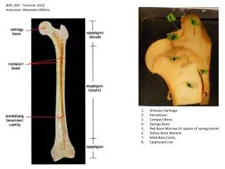
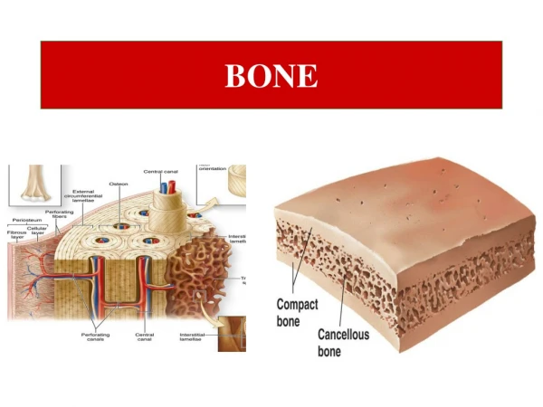
MORPHOLOGICAL DIFFERENTIATION OF CETACEAN BONE PARTICLES IN MEAT AND BONE MEAL M. Zadravec1*, Z. Kozarić2, S. Kužir2, M. Mitak1, M. Đuras2 1 Croatian Veterinary Institute 2 Faculty of Veterinary Medicine, University of Zagreb Introduction Meat and bone meal originating from terrestrial mammals, poultry and fish is conditionally prohibited for feeding fish, poultry and pigs and totally forbidden to ruminants because of the proven connection between the incidence of spongiform encephalopathy and animal feeding with feeds that contain meat and bone meal. The presence of bone particles originating from marine mammals in feed is not regulated by law, altough their carcasses can be involved in fish meal production. The aim of this study was to describe osteocyt lacunae of the bottlenose dolphin (Tursiops truncatus), as a representative of whales (Ordo: Cetacea). Further, our goal was to determine whether there are visible differences in the shape and size of osteocyte lacunae among dolphins, terrestrial mammals, poultry and fish. Materialsand methods The 5th right rib and the right humerus were sampled from 20bottlenose dolphins (Tursiops truncatus) (10 females, 10 males) ranging from less then one year to 21 years of age that had been found dead from 1990 to 2011 in the Croatian part of the Adriatic Sea.The rib was representing the axial and the humerus the appendicular skeleton hence any bone of the skeleton can occur in meat and bone meal.Each bone sample was separately crushed in a mortar to a particle size of less than 0.5 mm in order to resemble bone particles in meat and bone meal according to“The approval of alternative heat treatment systems for processing animal waste with a view to the inactivation of spongiform encephalopathy agents” (European Council 449/96). A small amount of bone particles was placed into Norland Optical Adhesive 65 (Norland Products , USA) that was dropped on micro slides and afterwards covered with cover glasses. The bone particles were analyzed under a magnification 200 x on Zeiss Axio Imager M2 microscope. Osteocyte lacunae density, shape of lacunae and position of canalliculae were analyzed for each sample. Length and width of osteocyte lacunae were measured and average values were calculated using Zeiss Axio Vision softwear. Results were compared with published values for fish, poultry, ruminant and pig (ARIES, 2004). Results cod (Clupea harengus) chicken (Gallus gallus) 5μm bottlenose doplphin (Tursiops truncatus) bovine (Bos taurus) pig (Sus domesticus) Table : The width and length of lacunae (* ARIES, 2004; ** this study) Conclusion Osteocyt lacunae of bottlenose dolphins are elliptical and evenly spaced through the bone fragment. They have clearly visible long but not dense canalicules. Hence bottlenose dolphin bone fragments can be differentiated well from fragments of bones of birds and pigs but not from ruminants in native microscopic preparations. For this reason we recommand additional analyzes such as enzyme-linked immunoassay or molecular methods. *Corresponding author: zadravec@veinst.hr