Download
1 / 65
650 likes | 832 Views
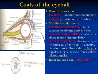
Three Coats of Eye Ball. 1.OUTER COAT 2.MIDDLE COAT 3.INNER COAT. Outer Coat. Tough Fibrous Coat Post 5/6 th of Globe White & Opaque Sclera Radius---12mm. Outer Coat. Tough Fibrous Coat Ant 1/6th of Globe Transparent Cornea Radius---8mm. Corneoscleral Limbus.

E N D
Three Coats of Eye Ball 1.OUTER COAT 2.MIDDLE COAT 3.INNER COAT
Outer Coat Tough Fibrous Coat Post 5/6th of Globe White & Opaque Sclera Radius---12mm
Outer Coat Tough Fibrous Coat Ant 1/6th of Globe Transparent Cornea Radius---8mm
Corneoscleral Limbus Junction of Cornea and Sclera Contains 1.Trabecular Meshwork 2.Canal of Schlemn
Size of Cornea Verticle-------10.6 mm Horizontal---11.7 mm Thickness Central portion----0.52 mm Peripheral portion----1 mm
Structure Three Layers 1. Epithelium & its Basement 2. Stroma & its ant condensation ( Bowman Zone) 3.Endothelium & its Basement (Descemet Membrane)
Structure From Anterior to Posterior 1. Epithelium 2. Bowman Zone 3. Stroma 4. Descemet Membrane 5. Endothelium
Epithelium • 50-60 µm thick • Covers the stroma anteriorly • Continuous with epithelium of conjunctiva • Life of epithelial cells is 7 days • Prevent aqueous solutions to penetrate
Epithelium • Surface cell layer • Wing cell layer • Basal cell layer • Basement membrane
Stroma 90% of the corneal thickness • Bowman Zone • Lamellar Stroma Once deformed its typical structure is not restored
Inner Lining • Descemet membrane (Regenerates) • Endothelium Single layer of cells Cells are tightly bound together Responsible for dehydration Never regenerates
Blood supply • Central cornea is avascular • Corneoscleral limbus is generously supplied by anterior conjuntival branches of the anterior ciliary arteries • Aqueous humor and tear film provides nutrients
Nerve Supply • Branches of the ophthalmic division of trigeminal nerve and are solely sensory • Most are concentrated in the anterior stroma beneath the Bowman zone and send branches forward into epithelium • Descemet membrane and endothelium are not innervated
Cornea • The microvilli of the anterior surface of the squamous cell layer are wet by the mucin of tear film • These cells are joined by tight junctions that exclude water soluble substances
Transparency • Tight junctions of the epithelial cells • Endothelial pump mechanism • Absence of blood vessels • Absence of pigments • Scarcity of cell nuclei in stroma • Regular structure of stroma
Signs of Corneal Disease Superficial 1.Punctate epithelial erosions Tiny ,slightly depressed, epithelial defects which stain with flourescein but not with rose Bengal PEE are non specific and may develop in a wide variety of keratopathies
Signs of Corneal Disease Superficial 2.Punctate epithelial keratitis It is the hallmark of viral infections. • Swollen epithelial cells • Visible unstained • Stains with rose bengal
Signs of Corneal Disease Superficial 3.Epithelial Oedema Sign of • Endothelial decompensation • Severe acute elevation of IOP
Signs of Corneal Disease Superficial 4.Filaments Small coma shaped mucus strands lined with epithelium. One end attached with epithelium
Signs of Corneal Disease Superficial 5.Pannus Inflammatory or degenerative ingrowth of fibro vascular tissue from limbus
Signs of Corneal Disease Stromal Lesions 1.Infiltrates Focal areas of active stromal inflammation 2. Oedema Increased corneal thickness Decreased transparency 3. Vascularization
Signs of Corneal Disease Lesions of Descemet Membrane • Breaks Corneal enlargement Keratoconus Birth trauma 2. Folds (Striate Keratopathy) Surgical trauma Ocular hypotony Stromal oedema
PRINCIPLES OF MANAGEMENT OF CORNEAL DISEASE • Control of infection • Control of inflammation • Promotion of re-epithelialization – lubrication – lid closure – bandage soft contact lens • Prevention of perforation – tissue adhesive glue – conjunctival flap – systemic immunosuppressive agents • Corneal grafting
MICROBIAL KERATITIS( Bacterial) • Ocular surface disease: Trauma, post-herpetic corneal disease, bullous keratopathy, corneal exposure, dry eye and diminished corneal sensation. • Contact lens wear
MICROBIAL KERATITIS( Bacterial) Pathogens which can produce corneal infection in intact epithelium. • 1.Neisseria gonorrhoeae • 2.Corynebacterium diphtheriae • 3.Listeria • 4.Haemophilus
Staph. aureus and strep. pneumoniae • Oval, yellow-white, densely opaque stromal suppuration surrounded by relatively clear cornea
Psuedomonas • Thick mucopurulent exudate, diffuse liquefactive necrosis and semi-opaque ground glass appearance of adjacent stroma
Enterobacteriaceae • Shallow ulceration, grey-white pleomorphic suppuration and diffuse stromal opalescence. Endotoxins may induce ring-shaped corneal infilterates
MANAGEMENT • History • Clinical examination (including staining and sensitivity) • Hospitalization • Corneal scrapping • Treatment
Treatment • Topical antibiotics – combination therapy with fortified amino glycoside and fortified cephalosporin or monotherapy with fluoroquinolone. Initial instillation at hourly intervals. • Subconjunctival injections • Systemic ciprofloxacin 750mg BD
Cycloplegics • Steroid therapy (controversial) • Corneal biopsy or excisional keratoplasty
Poor response to treatment • Wrong diagnosis • Wrong treatment • Drug toxicity
FUNGAL KERATITIS • Filamentous fungal keratitis –Aspergillus - Fusarium
History of vegetable matter injury • Greyish-white ulcer with indistinct margins • Surrounded by feathery infilterates • Ring infilterate • Endothelial plaque • Hypopyon
Candida keratitis • Usually develops in pre-existing corneal disease or immunocompromised patient • Yellow-white ulcer • Dense suppuration
D/D of fungal keratitis • Suppurative bacterial keratitis • Herpetic stromal necrotic keratitis
MANAGEMENT • Culture • Biopsy • Antifungal therapy – Initially broad-spectrum econazole 1% topically – Then depending upon sensitivity natamycin or imidazole for 6 weeks • Systemic ketoconazole • Therapeutic penetrating keratoplasty
ACANTHAMOEBA KERATITIS • Protozoan –active (trophozoite) –dormant (cystic) • Common in swimmers and CL wearers
CLINICAL FEATURES • Blurred vision and disproportionate pain • Patchy anterior stromal infilterates • Perineural infilterates (radial keratoneuritis) • Infilterates coalesce –ring abcess, ulceration and hypopyon • White satellite lesions
MANAGEMENT • Corneal scrappings stained with calcoflour white • Corneal biopsy • Treatment with chlorhexidine, polyhexamethylenebiguanide drops, dipropamidine and propamidine. • Therapeutic penetrating keratoplasty
HERPES SIMPLEX KERATITIS Primary ocular herpes: - Blepharoconjunctivitis - Keatitis (punctate epithelial)
DENDRITIC ULCER • Opaque cells arranged in a course punctate or stellate pattern • Central desquamation leads to a linear branching ulcer. –Fluorescein stain – Rose Bengal stain –Diminished corneal sensitivity • Anterior stromal infilterates • Geographical or amoeboid ulcer
Differential diagnosis • Herpes zoster keratitis • Healing corneal abrasion • Pseudodendrites due to soft contact lens • Acanthamoeba keratitis • Drug toxicity