Download
1 / 43
430 likes | 459 Views
Explore the intricate process of photosynthesis, where photoautotrophs like plants and bacteria capture sunlight energy to produce oxygen and glucose. Learn about the structure of chloroplasts, pigments involved in light absorption, and the two stages of photosynthesis. Delve into the mechanisms of light-dependent and light-independent reactions, electron transfer chains, and the production of ATP and NADPH. Discover the fascinating world of biochemical pathways and energy transformation in cells.

E N D
How Cells Acquire Energy Chapter 7
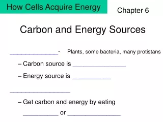
Photoautotrophs • Capture sunlight energy and use it to carry out photosynthesis • Plants • Some bacteria • Many protistans
Fig. 10-2 (a) Plants (c) Unicellular protist 10 µm (e) Purple sulfur bacteria 1.5 µm (b) Multicellular alga (d) Cyanobacteria 40 µm
Photosynthesis Energy-storing pathway Releases oxygen Requires carbon dioxide Aerobic Respiration Energy-releasing pathway Requires oxygen Releases carbon dioxide Linked Processes
Fig. 10-3 Leaf cross section Vein Mesophyll Stomata CO2 O2 Chloroplast Mesophyll cell Outer membrane Thylakoid Intermembrane space 5 µm Stroma Thylakoid space Granum Inner membrane 1 µm
Chloroplast Structure two outer membranes stroma inner membrane system (thylakoids connected by channels) Figure 7.3d, Page 116
Photosynthesis Equation LIGHT ENERGY 12H2O + 6CO2 6O2 + C2H12O6 + 6H2O Water Carbon Dioxide Oxygen Glucose Water In-text figurePage 115
Reactants 12H2O 6CO2 Products 6O2 C6H12O6 6H2O Where Atoms End Up In-text figurePage 116
Two Stages of Photosynthesis sunlight water uptake carbon dioxide uptake ATP ADP + Pi LIGHT-DEPENDENT REACTIONS LIGHT-INDEPENDENT REACTIONS NADPH NADP+ glucose P oxygen release new water In-text figurePage 117
Electromagnetic Spectrum Shortest Gamma rays wavelength X-rays UV radiation Visible light Infrared radiation Microwaves Longest Radio waves wavelength
Visible Light • Wavelengths humans perceive as different colors • Violet (380 nm) to red (750 nm) • Longer wavelengths, lower energy Figure 7.5aPage 118
Photons • Packets of light energy • Each type of photon has fixed amount of energy • Photons having most energy travel as shortest wavelength (blue-violet light)
Pigments • Color you see is the wavelengths not absorbed • Light-catching part of molecule often has alternating single and double bonds • These bonds contain electrons that are capable of being moved to higher energy levels by absorbing light
Fig. 10-7 Light Reflected light Chloroplast Absorbed light Granum Transmitted light
Variety of Pigments Chlorophylls a and b Carotenoids Xanthophils
Chlorophylls Main pigments in most photoautotrophs chlorophyll a Wavelength absorption (%) chlorophyll b Wavelength (nanometers) Figure 7.6a Page 119 Figure 7.7Page 120
Accessory Pigments Carotenoids, Phycobilins, Anthocyanins beta-carotene phycoerythrin (a phycobilin) percent of wavelengths absorbed wavelengths (nanometers)
Pigments in Photosynthesis • Bacteria • Pigments in plasma membranes • Plants • Pigments and proteins organized into photosystems that are embedded in thylakoid membrane system
Arrangement of Photosystems water-splitting complex thylakoid compartment H2O 2H + 1/2O2 P680 P700 acceptor acceptor pool of electron carriers PHOTOSYSTEM II stroma PHOTOSYSTEM I Figure 7.10Page 121
Light-Dependent Reactions • Pigments absorb light energy, give up e-, which enter electron transfer chains • Water molecules split, ATP and NADH form, and oxygen is released • Pigments that gave up electrons get replacements
Photosystem Function: Harvester Pigments • Most pigments in photosystem are harvester pigments • When excited by light energy, these pigments transfer energy to adjacent pigment molecules • Each transfer involves energy loss
Photosystem Function: Reaction Center • Energy is reduced to level that can be captured by molecule of chlorophyll a • This molecule (P700 or P680) is the reaction center of a photosystem • Reaction center accepts energy and donates electron to acceptor molecule
Fig. 10-12 STROMA Photosystem Photon Primary electron acceptor Light-harvesting complexes Reaction-center complex e– Thylakoid membrane Pigment molecules Special pair of chlorophyll a molecules Transfer of energy THYLAKOID SPACE (INTERIOR OF THYLAKOID)
Electron Transfer Chain • Adjacent to photosystem • Acceptor molecule donates electrons from reaction center • As electrons pass along chain, energy they release is used to produce ATP
Cyclic Electron Flow • Electrons • are donated by P700 in photosystem I to acceptor molecule • flow through electron transfer chain and back to P700 • Electron flow drives ATP formation • No NADPH is formed
Fig. 10-15 Primary acceptor Primary acceptor Fd Fd NADP+ + H+ Pq NADP+ reductase Cytochrome complex NADPH Pc Photosystem I ATP Photosystem II
Noncyclic Electron Flow • Two-step pathway for light absorption and electron excitation • Uses two photosystems: type I and type II • Produces ATP and NADPH • Involves photolysis - splitting of water
Machinery of Noncyclic Electron Flow H2O second electron transfer chain photolysis e– e– ATP SYNTHASE first electron transfer chain NADPH NADP+ ATP ADP + Pi PHOTOSYSTEM II PHOTOSYSTEM I Figure 7.13aPage 123
Fig. 10-13-5 Electron transport chain Primary acceptor Primary acceptor 4 7 Electron transport chain Fd e– Pq 2 e– 8 e– e– NADP+ + H+ H2O Cytochrome complex 2 H+ NADP+ reductase + 3 NADPH O2 1/2 Pc e– e– P700 5 P680 Light Light 1 6 6 ATP Pigment molecules Photosystem I (PS I) Photosystem II (PS II)
Energy Changes second transfer chain e– NADPH e– first transfer chain Potential to transfer energy (volts) e– e– (Photosystem I) (Photosystem II) 1/2O2 + 2H+ H2O Figure 7.13bPage 123
Chemiosmotic Model of ATP Formation • Electrical and H+ concentration gradients are created between thylakoid compartment and stroma • H+ flows down gradients into stroma through ATP synthesis • Flow of ions drives formation of ATP
Chemiosmotic Model for ATP Formation H+ is shunted across membrane by some components of the first electron transfer chain Gradients propel H+ through ATP synthases; ATP forms by phosphate-group transfer Photolysis in the thylakoid compartment splits water H2O e– acceptor ATP SYNTHASE ATP ADP + Pi PHOTOSYSTEM II Figure 7.15Page 124
Fig. 10-17 STROMA (low H+ concentration) Cytochrome complex Photosystem I Photosystem II Light 4 H+ NADP+ reductase Light 3 Fd NADP+ + H+ NADPH Pq Pc e– 2 e– H2O O2 1/2 1 THYLAKOID SPACE (high H+ concentration) 4 H+ +2 H+ To Calvin Cycle Thylakoid membrane ATP synthase STROMA (low H+ concentration) ADP + ATP P i H+
Light-Independent Reactions • Synthesis part of photosynthesis • Can proceed in the dark • Take place in the stroma • Calvin-Benson cycle
Overall reactants Carbon dioxide ATP NADPH Overall products Glucose ADP NADP+ Calvin-Benson Cycle Reaction pathway is cyclic and RuBP (ribulose bisphosphate) is regenerated
6 CO2 (from the air) Calvin- Benson Cycle CARBON FIXATION 6 6 RuBP unstable intermediate 12 PGA 6 ADP 12 ATP 6 ATP 12 NADPH 4 Pi 12 ADP 12 Pi 12 NADP+ 10 PGAL 12 PGAL 2 PGAL Pi P Figure 7.16Page 125 glucose
The C3 Pathway • In Calvin-Benson cycle, the first stable intermediate is a three-carbon PGA • Because the first intermediate has three carbons, the pathway is called the C3 pathway
Photorespiration in C3 Plants • On hot, dry days stomata close • Inside leaf • Oxygen levels rise • Carbon dioxide levels drop • Rubisco attaches RuBP to oxygen instead of carbon dioxide • Only one PGAL forms instead of two
C4 Plants • Carbon dioxide is fixed twice • In mesophyll cells, carbon dioxide is fixed to form four-carbon oxaloacetate • Oxaloacetate is transferred to bundle-sheath cells • Carbon dioxide is released and fixed again in Calvin-Benson cycle
CAM Plants • Carbon is fixed twice (in same cells) • Night • Carbon dioxide is fixed to form organic acids • Day • Carbon dioxide is released and fixed in Calvin-Benson cycle
Fig. 10-5-4 CO2 H2O Light NADP+ ADP + P i Calvin Cycle Light Reactions ATP NADPH Chloroplast [CH2O] (sugar) O2
Satellite Images Show Photosynthesis Atlantic Ocean Photosynthetic activity in spring Figure 7.20Page 128