Download
1 / 14
170 likes | 646 Views
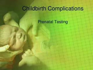
Childbirth Complications. Prenatal Testing. Fetoscopy.

E N D
Childbirth Complications Prenatal Testing
Fetoscopy • A fetoscopy is an endoscopic procedure (which means looking inside) during pregnancy to allow access to the fetus, the amniotic cavity, the umbilical cord, and the fetal side of the placenta. A small (3–4 mm) incision is made in the abdomen, and an endoscope (a device with a light and a small camera on the end of a long, flexible tube that is used to look inside a body cavity or organ) is inserted through the abdominal wall and uterus into the amniotic cavity. Fetoscopy allows physicians to examine the fetus before birth and it allows medical interventions to be performed. • Fetoscopy does pose a risk to the mother and baby so it is not utilized unless there is medical justification.
Ultrasound • This test can be done with an abdominal or vaginal probe depending on the stage of the pregnancy and what the doctor is looking for. The transducer or probe sends out high frequency sound waves which are sent into the body. As they pass through they bounce off different objects and are sent back as electrical signals, which are then processed and displayed as the image on the screen. • http://www.muschealth.com/video/Default.aspx?videoId=10092&cId=34&type=rel • http://www.mayoclinic.com/health/fetal-ultrasound/PR00139 • http://www.babycenter.com/2_ultrasound-exam_3658842.bc
Nuchal Translucency Screening Test (also called the NT or nuchal fold scan) • This prenatal test can help your healthcare practitioner assess your baby's risk of having Down Syndrome (DS) and some other chromosomal abnormalities as well as major congenital heart problems. • The NT test uses ultrasound to measure the clear (translucent) space in the tissue at the back of your developing baby's neck. Babies with abnormalities tend to accumulate more fluid at the back of their neck during the first trimester, causing this clear space to be larger than average. • The NT scan must be done between the 11th and 14th weeks of pregnancy. • It's usually offered along with an evaluation for the presence or absence of nasal bone, as well as a blood test, in what's known as first trimester combined screening. • First-trimester screening provides information about the baby's risk for chromosomal problems early in the pregnancy without subjecting the pregnancy to the risk of miscarriage from an invasive test like CVS.
Nuchal Translucency Screening Test • http://www.bing.com/videos/search?q=Nuchal+Scan&view=detail&mid=0A0101B7D03051462DCC0A0101B7D03051462DCC&first=0&FORM=NVPF • http://www.bing.com/videos/search?q=Nuchal+Scan&view=detail&mid=9C97B927287C84A3B12F9C97B927287C84A3B12F&first=0&FORM=NVPFVR
Amniocentesis • Then a long, slender needle is inserted through the skin of the abdomen into a safe location in the uterus and about 1 ounce of amniotic fluid is withdrawn. Ultra sound is used to guide the needle away from the baby and the placenta.
Amniocentesis (continued) • This test can be used to determine if a baby has a chromosomal disorder or for fetal lung maturity. • Amniocentesis is usually done between 15 and 18 weeks of gestation, although some practitioners are doing amniocentesis as early as 9 weeks. It normally takes two weeks to receive the results. The results can be very accurate, however, they cannot tell you the severity of a present defect. • There is a risk to the baby from this procedure. About 1 in 200 women will miscarry after the amniocentesis, even if the baby was unaffected, and about 1 in 1,000 will experience infection. • A laboratory analyzes the fluid sample, measuring the amount of alpha-fetoprotein (AFP) in the fluid. The lab also takes some of the baby's living cells from the fluid and allows them to reproduce for a week or two, then checks the cells for chromosomal abnormalities and evidence of certain genetic birth defects. • http://www.bing.com/videos/search?q=amniocentesis&view=detail&mid=C023CE167DF2E8378A0AC023CE167DF2E8378A0A&first=21&FORM=LKVR34&adlt=strict
Chorionic Villus Sampling (CVS) • Chorionic villi are tiny finger-shaped growths found in the placenta. The genetic material in chorionic villus cells is the same as that in the baby's cells. During CVS, a sample of the chorionic villus cells is taken and checked for problems. The procedure is generally done late in the first trimester, most often between the 10th and 12th weeks. • The chorionic villus sample can be collected by putting a thin flexible tube (catheter) through the vagina and cervix into the placenta. The sample can also be collected through a long, thin needle put through the belly into the placenta. Ultrasound is used to guide the catheter or needle into the correct spot for collecting the sample. http://www.muschealth.com/video/Default.aspx?videoId=10092&cId=34&type=rel
Chorionic Villus Sampling (continued) • CVS also increases the chance of: • Developing a uterine infection. • Having a miscarriage. The chance of miscarriage is higher for transcervical CVS than for abdominal CVS. Overall, one study showed the risk of miscarriage from CVS is about 1 in 100 when done by a highly trained provider. • Having a baby with arm or leg abnormalities though the chance of this happening is very low, especially when the test is done after 10 weeks.
Glucose Screening Test (Glucose Tolerance Test) • A positive result doesn't always mean a pregnant woman has gestational diabetes. Only about a third of women who test positive on the glucose screen actually have the condition. • Between 2 and 5 percent of expectant mothers develop gestational diabetes, making it one of the most common health problems during pregnancy. • Most doctors routinely recommend a glucose screening test between 24 and 28 weeks of pregnancy. • The pregnant woman is given a sugar solution that contains 50 grams of glucose. It tastes like a very sweet soda pop (and comes in several flavors). The pregnant woman must get all of the solution down within five minutes. An hour later a blood sample from your arm will be taken and analyzed to check blood sugar levels.
Alpha-fetoprotein (AFP) • The alpha-fetoprotein test (AFP test) involves drawing a blood sample from the mother to check the levels of AFP. AFP is a protein secreted by the fetal liver and excreted (eliminated) in the mother's blood. It is generally used to provide a screening for neural tube defects like spina bifida and ancephaly. It can also indicate: abdominal wall defects, esophageal and duodenal atresia, some renal and urinary tract anomalies, Turner syndrome, some low birth-weight fetuses, placental complications, and the presence of Down Syndrome.
Triple Screening/Maternal Serum Screening (MSS) • The triple screen, also known as a maternal serum screening test or MSS, is a simple and completely safe blood test that measures not only AFP, but hCG and estriol as well. These are all hormones produced by the fetus and passed into the mother’s bloodstream. The test is performed between the 15th and 18th weeks, with the results usually available within one week. Elevated levels of AFP in the mother’s blood can indicate a neural tube defect in the baby such as a deformity of the spinal canal known as spina bifida. Lower levels of AFP suggest a possible child with Down syndrome or other chromosomal defect. • This test is more accurate than the AFP test and screens for additional genetic problems, and is beginning to replace the standard AFP. Generally speaking, any combination of the testing will identify 60% of the babies with Down Syndrome and 80-90% of the babies with neural tube defects. The AFP test is generally most sensitive between the 15th and 17th weeks of pregnancy, while the triple screen can be done a bit earlier.
Quadruple Screening • A fairly recent addition to the screening tests, the quadruple test is almost identical to the triple screen, except that it tests for one more marker. Again using a sample of the mother's blood, a quadruple screen measures a woman's levels of alpha fetoprotein, hCG, estriol and inhibin to indicate her baby's risk of Downs syndrome. The quadruple test is about 81% effective and is usually performed around the 16th week of pregnancy. If a risk of Downs syndrome is indicated, amniocentesis can be performed to confirm the diagnosis.
Problems with screening tests • The most common risk of having a triple or quadruple screen test is unnecessary worry. Most women have normal serum screen results. Of women who have positive results, most turn out to have no problems. An incorrect fetal age or pregnancy with twins can lead to a false-positive result. The quadruple screen may be slightly less likely to give you a false-positive result. Positive results are normally followed with fetal ultrasound or amniocentesis to see if the baby really has a birth defect.