IV INFUSION
370 likes | 824 Views
Intravenous (IV) therapy is a medical procedure that delivers liquids directly into a vein, which can be done continuously or intermittently. It's the fastest route for administering fluids and medications, making it essential in emergency care and routine treatments. The most common method is through peripheral IV lines, which are inserted into arm or hand veins. Various fluids are utilized, including crystalloids and colloids, with normal saline being a primary choice. However, IV therapy carries risks such as infections and fluid overload that must be managed carefully.

IV INFUSION
E N D
Presentation Transcript
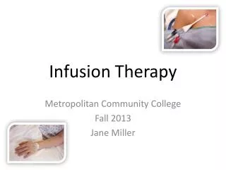
Intravenous therapy Intravenous therapy or IV therapy is the administration of liquid substances directly into a vein. It can be intermittent or continuous; continuous administration is called an intravenous drip. The word intravenous simply means "within a vein", but is most commonly used to refer to IV therapy Compared with other routes of administration, the intravenous route is the fastest way to deliver fluids and medications throughout the body. Some medications, as well as blood transfusions and lethal injections, can only be given intravenously
Peripheral IV lines This is the most common intravenous access method in both hospitals and paramedic services. A peripheral IV line consists of a short catheter (a few centimeters long) inserted through the skin into a peripheral vein. A peripheral vein is any vein that is not in the chest or abdomen. Arm and hand veins are typically used although leg and foot veins are occasionally used. On infants the scalp veins are sometimes used.
Peripheral IV lines Part of the catheter remains outside the skin, with a hub that can be connected to a syringe or an intravenous infusion line,. The caliber of cannulae is commonly indicated in gauge, with 14 being a very large cannula (used in resuscitation settings) and 24-26 the smallest. The most common sizes are 16-gauge (midsize line used for blood donation and transfusion), 18- and 20-gauge (all-purpose line for infusions and blood draws), and 22-gauge (all-purpose pediatric line). 12 and 14-gauge peripheral lines actually deliver equivalent volumes of fluid faster than central lines, accounting for their popularity in emergency medicine; these lines are frequently called "widebores" or "trauma lines
IV fluids Crystalloids are aqueous solutions of mineral salts or other water-soluble molecules. Colloids contain larger insoluble molecules, such as gelatin; blood itself is a colloid The most commonly used crystalloid fluid is normal saline, a solution of sodium chloride at 0.9% concentration, which is close to the concentration in the blood (isotonic). Ringer's lactate or Ringer's acetate (ASERING) is another isotonic solution often used for large-volume fluid replacement. A solution of 5% dextrose in water, sometimes called D5W, is often used instead if the patient is at risk for having low blood sugar or high sodium. The choice of fluids may also depend on the chemical properties of the medications being given.
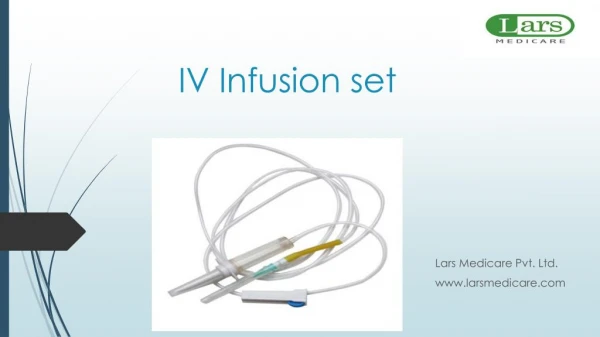
Intravenous fluids must always be sterile A standard IV infusion set consists of a pre-filled, sterile container (glass bottle, plastic bottle or plastic bag) of fluids with an attached drip chamber which allows the fluid to flow one drop at a time, making it easy to see the flow rate (and also reducing air bubbles); a long sterile tube with a clamp to regulate or stop the flow; a connector to attach to the access device; and connectors to allow "piggybacking" of another infusion set onto the same line, e.g., adding a dose of antibiotics to a continuous fluid drip
An infusion pump allows precise control over the flow rate and total amount delivered, but in cases where a change in the flow rate would not have serious consequences, or if pumps are not available, the drip is often left to flow simply by placing the bag above the level of the patient and using the clamp to regulate the rate; this is a gravity drip
Intermittent infusion Intermittent infusion is used when a patient requires medications only at certain times, and does not require additional fluid. It can use the same techniques as an intravenous drip (pump or gravity drip), but after the complete dose of medication has been given, the tubing is disconnected from the IV access device. Some medications are also given by IV push, meaning that a syringe is connected to the IV access device and the medication is injected directly (slowly, if it might irritate the vein or cause a too-rapid effect). Once a medicine has been injected into the fluid stream of the IV tubing there must be some means of ensuring that it gets from the tubing to the patient. Usually this is accomplished by allowing the fluid stream to flow normally and therby carry the medicine into the bloodstream; however, a second fluid injection is sometimes used, a "flush", following the injection to push the medicine into the bloodstream more quickly.
Risks of intravenous therapy Infection Phlebitis Infiltration This occurs when the tip of the IV catheter withdraws from the vein or pokes through the vein into surrounding tissue, or when the vein's wall becomes permeable and leaks fluid (in this instance it is said that the cannula has 'tissued'). It occurs frequently with peripheral IVs, and requires replacement of the IV at a different location.
Risks of intravenous therapy Fluid overload This occurs when fluids are given at a higher rate or in a larger volume than the system can absorb or excrete. Possible consequences include hypertension, heart failure, and pulmonary edema Electrolyte imbalance Administering a too-dilute or too-concentrated solution can disrupt the patient's balance of sodium, potassium, and other electrolytes. Hospital patients usually receive blood tests to monitor these levels. Embolism
USES For diagnostic purposes Therapeutic(excess iron or erythrocytes) For later purposes(blood transfusion)
EQUIPMENTS Evacuated tube system (in developed countries) OR Needle & syringe
PROCEDURE Assemble equipment Prepare the patient Select the site Perform hand hygiene and put on gloves Disinfect the entry site Take blood Fill the laboratory sample tubes
Draw blood as follows… Anchor the vein by holding the patient's arm and placing a thumb below the venepuncture site. Ask the patient to form a fist so the veins are more prominent. Enter the vein swiftly at a 30 degree angle or less, and continue to introduce the needle along the vein at the easiest angle of entry. Once sufficient blood has been collected, release the tourniquet before withdrawing the needle. Withdraw the needle gently and apply gentle pressure to the site with a clean gauze or dry cotton-wool ball. Ask the patient not to bend the arm, because doing so causes a haematoma.
COMPLICATIONS Hematoma Infection Nerve damage Extravasation Syncope & fainting Petechiae Excessive bleeding Edema Thrombosis Allergies