Understanding the Cardiovascular System: Anatomy and Functions
260 likes | 360 Views
This lecture provides a comprehensive overview of the cardiovascular system, focusing on the heart, blood vessels, and their functions. Students will learn to identify the components, including the heart's structure—its chambers, valves, and the vascular network of arteries, veins, and capillaries. Key topics include the portal system, sinusoids, arteriovenous anastomosis, and end-arteries. By the end, students will grasp how this vital system transports blood, oxygen, nutrients, and waste, crucial for maintaining body homeostasis.

Understanding the Cardiovascular System: Anatomy and Functions
E N D
Presentation Transcript
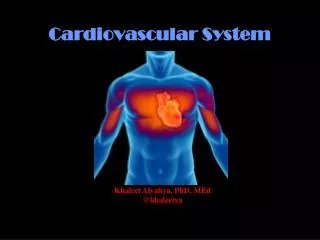
Cardiovascular System Khaleel Alyahya, PhD, MEd @khaleelya
OBJECTIVES At the end of the lecture, students should be able to: • Identify the components of the cardiovascular system. • Describe the Heart in regard to (position, chambers and valves). • Describe the Blood vessels (Arteries, Veins and Capillaries). • Describe the Portal System. • Describe the Sinusoids. • Describe the Functional and Anatomical end arteries. • Describe the Arteriovenous Anastomosis.
Pump: HEART Network of Tubes: BLOOD VESSELS CONTENT
It is a transportation system which uses the blood as the transport vehicle. It carries oxygen, nutrients, cell wastes, hormones and many other substances vital for body homeostasis. Provide forces to move the blood around the body by the beating Heart. FUNCTIONS
It is a hollow, cone shaped muscular pump that keeps circulation going on. It is the size of hand’s fist of the same person. It has: Apex Base Surfaces: Diaphragmatic & Sternocostal Borders: Right, Left, Inferior. THE HEART
It lies in a centrally located partition in the thoracic cavity known as the Middle Mediastinum between the two pleural sacs. Enclosed by a double sac of serous membrane (Pericardium). 2/3 of the heart lies to the left of median plane. LOCATION OF THE HEART
ATRIA: They bare two (Right & Left). Superior in position. They are the receiving chambers. They have thin walls. The upper part of each atrium is the Auricle. The Right Atrium receives the venous blood coming to the heart. Left Atrium receives arterial blood coming from the lungs. CHAMBERS OF THE HEART
VENTRICLES: The inferior chambers. They are two (right & left). They have thick walls. They are the discharging chambers (actual pumps). Their contraction propels blood out of the heart into the circulation. CHAMBERS OF THE HEART
The heart has Four Valves: Two Atrioventricular valves. One Aortic Semilunar valve. One Pulmonary Semilunar valve. VALVES OF THE HEART
Atrioventricular Valves: Valves between atria & ventricles. They allow the blood to flow in one direction from the atria to the ventricles. Right AVV (Tricuspid). Left AVV (Bicuspid). VALVES OF THE HEART
Semilunar Valves (Aortic & Pulmonary): Between the right and left ventricles and the great arteries leaving the heart. They allow the flow of blood from the ventricles to these arteries. VALVES OF THE HEART
Arteries: Thick walls. Do not have valves. The smallest arteries are arterioles. Veins: Thin walls. Many of them possess valves. The smallest veins are venules. Capillaries Connect arterioles and venules. Help to enable the exchange of water, oxygen and other nutrients between blood and the tissues. BLOOD VESSELS
They transport blood from the heart and distribute it to the various tissues of the body through their branches. Carry oxygenated blood away from the heart. two exceptions: the pulmonary and the umbilical arteries ARTERIES
It is the connection of two structures It is the joining of terminal branches of the arteries. ANASTOMOSIS
It is the artery that is the only supply of oxygenated blood to a portion of tissue. splenic artery & renal artery Two types of end arteries are: Anatomic (True) End Artery No anastomosis Functional End Artery Ineffectual anastomosis END ARTERIES
They transport blood back to the heart. The smaller veins (Tributries) unite to form larger veins which commonly join with one another to form Venous Plexuses. Carry deoxygenated blood toward the heart. two exceptions: the pulmonary and the umbilical veins VEINS
Two veins that accompany medium sized deep arteries Vena comitans is Latin for accompanying vein. They are found in close to arteries so that the pulsations of the artery aid venous return. Venae comitantes are usually found with smaller arteries, especially those in the extremities. Larger arteries do not have venae comitantes. They usually have a single, similarly sized vein. DEEP VEINS (VENAE COMITANTES)
Microscopic vessels in the form of a network. They connect the Arterioles to the Venules. they help to enable the exchange of water, oxygen and many other nutrients between bloodand the tissues CAPILLARIES
Direct connections between the arteries and veins without the intervention of capillaries. Found in tips of the fingers and toes. ARTERIOVENOUS ANASTOMOSIS
It is a system of vessels interposed between two capillary beds. Veins leaving the gastrointestinal tract do not go direct to the heart. They pass to the Portal Vein. This vein enters the liver and breaks up again into veins of diminishing size which ultimately join capillary like vessels (Sinusoids). PORTAL CIRCULATION SYSTEM
Thin walled blood vessels like capillaries. They are wider with irregular cross diameter. They are found in: Liver Spleen Bone marrow Some endocrine glands SINUSOIDS
The cardiovascular system is a transporting system. It is composed of the heart and blood vessels. The heart is cone shaped, covered by pericardium and composed of four chambers. The blood vessels are the arteries, veins and capillaries. Arteries transport the blood from the heart. The terminal branches of the arteries can anastomose with each other freely or be anatomic or functional end arteries. Veins transport blood back to the heart. Capillaries connect the arteries to the veins. Sinusoids are special type of capillaries. The portal system is composed of two sets of capillaries. The veins from the GIT go first to the liver through the portal vein. SUMMARY
Review Question # 1 • Which one of the following is NOT true? • Right atria receive blood from the body. • The valve between right atrium and right ventricle called “Bicuspid”. • Left ventricle discharging blood to the body. • Right ventricle receives blood from right atrium. • Valves allow blood to move one way only.
Review Question # 2 • Which statement of the following is TRUE? • Arteries transport blood from the heart to the body. • Arteriovanous anastomosis found in tips of the fingers and toes. • Capillaries connect the Arterioles to the Venules. • Anastomosis is the joining of terminal branches of the arteries. • Veins leaving the gastrointestinal tract do not go direct to the heart.


