
Glaucoma
E N D
Presentation Transcript
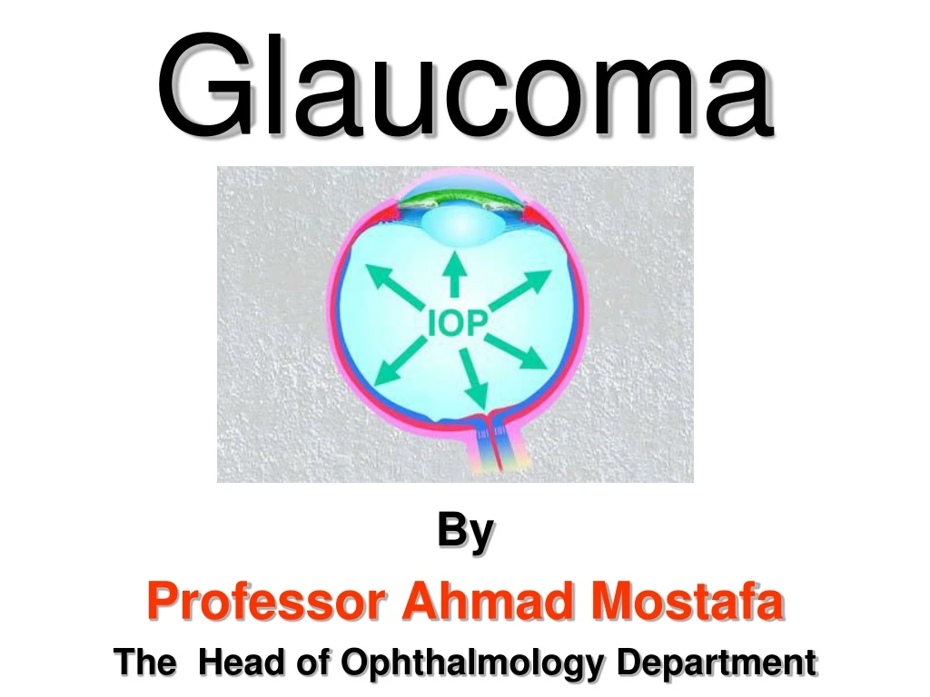
Glaucoma By Professor Ahmad Mostafa The Head of Ophthalmology Department
►The following will be discussed in the same order: I. APPLIED ANATOMY & PHYSIOLOGY II. GLAUCOMA: ● Definition of Glaucoma ● Classification of Glaucomas ● Clinical Picture of glaucomas ● Treatment of Glaucoma: ● Medical therapy of Glaucoma ● Laser therapy in Glaucoma ● Glaucoma Surgery
APPLIED ANATOMY & PHYSIOLOGY
I. Applied Anatomy and Physiology • Anatomy of the Angle of the anterior chamber • Physiology of Aqueous: Secretion & outflow • Intraocular Pressure (IOP)
Normal Angle Angle Closure
(b) Physiology of Aqueous: Secretion & outflow 1. Aqueous secretion • Active secretion accounts for approximately 80% of aqueous production. The aqueous is secreted by the non-pigmented ciliary epithelium via an active metabolic process b. Passive secretion accounts for the remaining 20%. Here, aqueous is produced by passive processes such as ultrafiltration and and diffusion which is dependant on the level of blood pressure in ciliary capillaries, the plasma oncotic pressure, and the level of intraocular pressure (IOP).
2. Aqueous outflow Aqueous flowsfrom the PC into the AC through the pupil and is drained by the following 2 routes: 1. The trabecular route(90% of aqueous outflow), is through the trabeculum into Schlemm's canal. It is then drained by the episcleral veins (a). 2. The uveoscleral route(10% of aqueous outflow). The aqueous passes across the ciliary body into the suprachoroidal space and is drained by the venous circulation in the ciliary body, choroid and sclera (b). N.B.Some aqueous also drains via the iris(c). • Normal aqueous outflow: • Trabecular route • (b) uveoscleral route • (c) through the iris
What is the normal IOP? • Factors affecting the IOP • Fluctuations of IOPoccur with the time of the day (diurnal variation),heartbeat, blood pressure, and respiration. • The pattern of diurnal variation differs in normal than glaucomatous eyes in that the difference between the morning and evening readings of IOP does not exceed 4 mmHg in normal eyes, but it may reach 10 mmHg or more in glaucomatous eyes.
Measurement of IOP: • Digital • Indentation Tonometry • Applanation Tonometry • Perkins Tonometer
Indentation Tonometry Applanation Tonometry
Indentation Tonometry Applanation Tonometry Advantages of applanation over indentation tonometry
II. Classification of Glaucoma ►There are many systems for classification of glaucoma: • According to the onset into: a. Congenital glaucoma b. Infantile glaucoma c. Juvenile glaucoma d. Adult glaucoma
2. According to the state of the drainage angle into: 1. Open-angle glaucoma (OAG) 2. Angle-closure glaucoma (ACG) 3. According to the presence or absence of associated factors contributing to the rise of IOP into: A. Primary glaucomas: ● OAG ● ACG ● Congenital B. Secondary glaucomas: ● OAG ● ACG ● Congenital
However, for the purpose of discussion, glaucoma can be classified into: • Congenital Glaucomas: 1. 1ry congenital glaucoma 2. 2ry congenital glaucoma B.Adult Glaucomas: (I) Primary Glaucomas i. 1ry OAG ii. 1ry ACG (II) Secondary Glaucomas i. 2ry OAG ii. 2ry ACG
Clinical Types of Glaucoma
I.Primary Congenital Glaucoma Buphthalmos
Primary Congenital Glaucoma(Buphthalmos) ● Definition: • A condition in which elevation of IOP is caused by a congenital maldevelopment of the trabeculum and the iridotrabecular junction, not associated with other major ocular anomalies (isolated trabeculodysgenesis). Bilateral Buphthalmos L Buphthalmos
Clinical Picture: • Symptoms: Photophobia and lacrimation
B. Signs: • Cornea: - Progressive enlargment - Edema (hazy) - Haab’s Striae (rupture of DM) - Opacities (late) 2. Sclera: blue (streached & thinned) 3. AC: Deep 4. Lens: may be subluxated 5. Fundus: Glaucomatous cupping
● Differential Diagnosis of Buphthalmos: 1◘ Cloudy cornea at birth ▪ Trauma ▪ Intrauterine rubella infection ▪ Peter's anomaly ▪ Metabolic disorders (e.g.mucopolysaccharidoses)
2◘ Large cornea at birth: ▪ Megalocornea ▪ High myopia Megalocornea 3◘ Lacrimationmay be confused with congenital epiphora (delayed canalization of the NLD). 4◘ Secondary glaucomas in infants . Tumors (e.g. retinoblastoma) . Persistant hyperplastic primary vitreous (PHPV) . Advanced retinopathy of prematurity (ROP)
Treatment • Goniotomyis the procedure of choice for early cases in where the angle structures can be seen. (2)Trabeculectomy is useful if the cornea is cloudy or if goniotomy fails. (3) Use of glaucoma filtering shunts for advaned & recurrent cases (4) Treat refractive errors and amblyopia.