Sad1
Fig. S1 Itadani et al. WT. cam1-22,117. Sad1. merge. Sad1. merge. Cut12-GFP. merge. Cut12-GFP. merge. Alp4-GFP. merge. Alp4-GFP. merge. Sid4-GFP. Sid4-GFP. merge. merge. merge. Pcp1-GFP. Pcp1-GFP. merge.

Sad1
E N D
Presentation Transcript
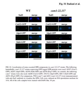
Fig. S1 Itadani et al. WT cam1-22,117 Sad1 merge Sad1 merge Cut12-GFP merge Cut12-GFP merge Alp4-GFP merge Alp4-GFP merge Sid4-GFP Sid4-GFP merge merge merge Pcp1-GFP Pcp1-GFP merge FIG. S1. Localization of some essential SPB components in cam1-22,117 strains. The following homothallic cam1-22,117 strains expressing GFP-tagged SPB proteins were used: AI197 (Cut12-GFP), AI207 (Alp4-GFP), AI190 (Sid4-GFP) and AI350 (Pcp1-GFP). As controls, the following cam1+ strains were also used: AI200 (Cut12-GFP), YN372 (Alp4-GFP), S60-3 (Sid4-GFP) and AI351 (Pcp1-GFP). For comparison, TN8 (cam1+) and AI22 (cam1-22,117)were immunostained with anti-Sad1 antibody to visualize the SPB. Cells were cultured on SSA sporulation medium for 18 h. All of the cells sampled were stained with DAPI. Bar, 10 m.