Download
1 / 17
170 likes | 408 Views
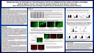
BRAIN DELIVERY OF PROTEINS BY THE INTRANASAL ROUTE OF ADMINISTRATION USING CATIONIC LIPOSOMES. Presented by Mattia M. Migliore February 23, 2007 Graduate Materials Links Symposium Northeastern University, Boston MA 02115. Introduction :.

E N D
BRAIN DELIVERY OF PROTEINS BY THE INTRANASAL ROUTE OF ADMINISTRATION USING CATIONIC LIPOSOMES Presented by Mattia M. Migliore February 23, 2007 Graduate Materials Links Symposium Northeastern University, Boston MA 02115
Introduction: • Neurodegenerative diseases cause the progressive destruction of either peripheral or central nervous system neurons and result in significant cognitive and/or motor dysfunction. • Include Alzheimer’s disease, Parkinson’s disease, ALS, brain cancer, Huntington’s disease, and multiple sclerosis. • Neurotrophic factors are growth factors that stimulate neuronal regeneration and/or prevent neuronal cell death.
Introduction(cont.): • The clinical use of neurotrophic factors and other therapeutic proteins has been limited due to their inability to cross the blood-brain barrier. • Currently, administration is limited to invasive intracerebral infusions. • The purpose of the present study was to develop a cationic liposomal system for nasal delivery of proteins to the brain.
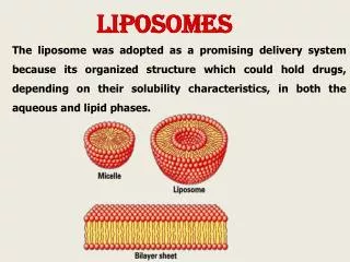
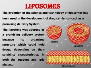
Rationale: • The intranasal route of administration was selected because it can bypass the blood-brain barrier, avoids systemic absorption, and limits potential peripheral side effects. • Two different preparations of cationic liposomes were generated containing a fluorescently tagged model protein, Alexa-488 ovalbumin. • Ovalbumin (OVAL) was selected because its molecular weight (45KDa) is similar to the molecular weight of several neurotrophic factors.
Specific Aims: • Aim # 1: To characterize and optimize a nanoparticle formulation for intranasal delivery of OVAL. • Aim # 2: To determine brain delivery, both qualitatively and quantitatively. • Aim # 3: To determine brain distribution of OVAL, and protein integrity. • Aim # 4: To determine co-localization of OVAL with a dopamine neuronal marker, tyrosine hydroxylase.
Specific Aim # 1: Data presented as mean ± SEM
Specific Aims # 2: Qualitative Determination of Protein Brain Delivery: Intranasal Alexa 488-OVAL (no nanoparticles) 24 hr time point, striatum(20x) Intranasal Alexa 488-OVAL (Preparation # 1) 24 hr time point, striatum (20x) Intranasal Alexa 488-OVAL (Preparation # 2) 24 hr time point, SN (20x)
Specific Aim # 2: Quantitative Analysis of Protein Brain Delivery: Time course of brain uptake of 111In-OVAL (1 µg/ µl) for liposomal preparations or control (PBS)
Biodistribution Study: 111In OVAL in Preparation #2 (1 µg/µl) 111In OVAL in Preparation #2 (2 µg/µl)
Time course of brain uptake of 111In-OVAL (2 µg/µl) for liposomal Preparation # 2 or control (PBS)
Specific Aim # 3: Protein Brain Distribution Localization of Alexa 488-OVAL in the corpus striatum after intranasal administration. Alexa 488-OVAL (Preparation # 1), 24 hr time point (20x). Intracellular uptake of Alexa 488-OVAL in the substantia nigra after intranasal administration. Alexa 488-OVAL (Preparation # 2), 24 hr time point (40x).
Posterior Alexa-488 ovalbumin brain distribution Midbrain Striatum Olfactory bulb Frontal
Specific Aim # 3: ProteinIntegrity Intranasal OVAL (Preparation # 2, 2 μg/μl) 24 hr time point, SN (40x). Intranasal OVAL (Preparation # 2, 2 μg/μl) 1 hr time point, striatum (20x).
Specific Aim # 4: Co-localization of Alexa-488 OVAL with Tyrosine Hydroxylase Intranasal Alexa 488-OVAL (Preparation # 2) 24 hr time point, SN (40x) TH positive dopamine neurons SN (40x) Merged image
Conclusions: • Liposomal preparations of OVAL effectively deliver the protein to brain after intranasal administration to rats. • The highest brain levels were detected at the shortest time point, i.e. 1 hr after administration. • Liposomal preparations increase brain residence time of the protein at the 24 hr time point when compared to control. • Liposomal OVAL delivered intranasally yields discrete protein deposits in both striatum and SN, with apparent cellular uptake in the SN by 24 hrs. • Intranasal administration of the 2 µg/µl form of liposomal Preparation #2 provides higher brain levels and reduced distribution to the GI tract relative to the more dilute form.
Work was supported by the NCI-NSF IGERT Nanomedicine S&T award, and a 2005 AFPE (American Foundation for Pharmaceutical Education) fellowship. Thesis committee: Dr. Barbara Waszczak Dr. Mansoor Amiji Dr. Robert Campbell Dr. Rebecca carrier Dr. Ralph Loring Dr. Robert Schatz Dr. Tushar Vyas Dr. Amiji’s lab: Sandip Lillian Mayank Dr. Campbell’s lab: Suman and Ashish. Dr. William Hartner. Dr. Torchilin’s lab. Eunice. Fran. Acknowledgements: