Download
1 / 97
1.02k likes | 1.43k Views
Paediatric Cardiac disorders. Robyn Smith Department of Physiotherapy, UFS, 2011.

E N D
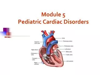
Paediatric Cardiac disorders Robyn Smith Department of Physiotherapy, UFS, 2011
Dealing with a child with cardiac dysfunction is often disconcerting and we are often unsure of how to proceed this lecture aims to provide an overview of common heart pathology in children and the physiotherapy management thereof
Background • Congenital heart defects (CHD) occur in 1% of the live births (6 of every 1 000) • Most common congenital abnormality seen • Approximately 1/3 of these children will require surgery, whilst the rest of the cases resolve spontaneously or are deemed haemodynamically insignificant • Early surgical intervention is recommended to limit CVS and neurodevelopmental complications. Most children are operated on before 1 year of age
Background • Mortality for children with CHD has decreased significantly ( ≤ 5 %) as a result of medical and surgical advances, and many of these children are surviving well into adulthood. • The decreasing mortality rates has resulted in the shift in focus to the neurodevelopmental status of these children and ways of addressing the associated developmental delays
Background • As PT’s we will encounter children with CHD in all clinical settings we work in • Acute care setting – pre/postoperatively • Sub-acute care setting in the ward • Out patient department • As PT’s we need to know: • What CHD is • Types of cardiac disorders • How the child’s CVS system is affected during exercise • Prevalent complications associated CHD
Aetiology • In most cases of CHD the aetiology is multi-factorial and include • genetic inheritance (patterns not yet clear) • Maternal conditions • Environmental factors • Above factors interact during the first 8-10 weeks of gestation a critical development phase of the heart
Normal foetal circulation • Foetal heart in not dependant on the lungs for respiration. Instead the placenta is used for gaseous exchange. • The R and L ventricles exist in a parallel circuit • Blood travels through the umbilical vein through the ductusvenosus to the foetal heart via the IVC to the RA and through the foramen ovale to the LA • The SVC leads to the RA to the RV to the pulmonary artery to the lungs or ductusarteriosus bypassing the lungs into the descending aorta to perfuse the lower extremities and the body, travelling back to the placenta via the umbilical arteries.
Normal foetal circulation • The blood travelling through the left ventricle to the aorta perfuses the upper extremities and the brain. • All of the blood flowing through the chambers of the heart, arteries and veins is rich in Oxygen • The vessels for pulmonary circulation in the foetus are vasoconstricted. All blood travelling in the arteries to the lungs is oxygen rich and contributes to the nourishment of the lung tissue
Changes in the circulatory system at birth • As the baby takes its first breath the lungs expand, causing the lung P to fall. This allows the blood to move more easily into the lung. • After reaching the lungs and being oxygenated the blood is moved to the LA. The P on the L side of the atrial septum becomes higher than on the R causing the foramen ovale to gradually close (closed by 3/12) • Once the lungs are filled with air and the oxygen level in the child’s blood rises the muscle wall of the ductusarteriosus contracts no longer allowing blood to flow through the ductus. The ductusarteriosus closes 10-15 hours after birth. • Now child has separate oxygenated and de-oxygenated blood and relies fully on the lungs for gaseous exchange
Congenital heart defects • At any point in the development of the cardiac system problems can arise leading to congenital heart disease. • CHD can be classified into two main groups: • Cyanotic lesions ( ↓O2 saturation in the blood) • Acyanotic lesions (O2 saturation unaltered, but can result in pressure or volume related issued)
Classification of Acyanoticheart lesions • Coarctation of aorta • Pulmonary stenosis obstructive in nature • Aortic stenosis • Patent ductusarteriousus • Atrialseptal defects • Ventral septal defects increased pulmonary bloodflow • Atrioventricularseptal defects with shunting O2 rich blood from left to right “PINK BABY”
Patent DuctusArteriosus (PDA) • The ductusarteriousus is the foetal vascular connection between the main pulmonary trunk and the aorta which under normal circumstances closes soon after birth (usually within the first week of life). • If it stays open excessive blood shunts from the aorta ton the lungs • Causing pulmonary oedema and in the long run pulmonary vascular disease • Symptoms may vary from mild to severe depending on the magnitude of the shunt • Very common in premature infants and may further complicate weaning from the ventilator and result in CHF
Patent DuctusArteriosus (PDA) • clinical signs and symptoms of significant PDA • Poor feeding • Failure to thrive (below weight for and height for age) • Sweating with crying or play • Persistent tachypnoea or breathlessness (dyspnoea) • Easy tiring • Tachycardia • Frequent lung infections • A bluish or dusky skin tone • Developmental delay
Patent DuctusArteriosus (PDA) • Management Closing of PDA can be induced using medication (indomethacin) Surgically • Surgical correction is done via a thoracotomy
AtrialSeptal Defect (ASD) • An ASD is an opening or whole in the wall separating the atria • This permits free communication of blood between the two atria. • Seen in 10% of all congenital heart disease • Rarely presents with signs of congestive heart failure or other cardiovascular symptom • Most are asymptomatic but may have easy fatigability or mild growth failure. The right atrium and ventricle may enlarge over time • Cyanosis does not occur unless pulmonary hypertension is present.
AtrialSeptal Defect (ASD) Management: • Surgical or catheterization closure is usually indicated • Closure is performed electively between ages 2 & 5 yrs if the whole has not closed in order to avoid late complications. Children may be on anticoagulant therapy for 6 months to prevent clotting • Surgical correction is done earlier in children with congestive heart failure or significant pulmonary hypertension
VenticularSeptal Defect (VSD) • A VSD is an abnormal opening in the ventricular septum, which allows free communication between the right and left ventricles ventricles. • Oxygen rich blood in the left ventricle is then pumped into the right ventricle through the opening instead of to the body. In a large VSD excessive blood is pumped to the lungs resulting in congestion and shortness of breath. • In return excessive amounts of blood are pumped back from the lungs to the left heart overburdening and enlarging it resulting in CHF
VenticularSeptal Defect (VSD) • In case of a small VSD most children are asymptomatic and 50% will close spontaneously by age 2yrs • In the case of a moderate or large VSD the child will be symptomatic. This may include dyspnoea, feeding difficulties, failure to thrive recurrent respiratory infections and profuse sweating
VenticularSeptal Defect (VSD) Management • In case of a small VSD 50% will close spontaneously by age 2yrs • Large VSD’s are usually closed surgically
AtrioventricularSeptal Defect (AVSD) • AVSD results from the incomplete fusion of the tendocardial cushions, which help to form the lower portion of the atrial septum, the membranous portion of the ventricular septum and the septal leaflets of the triscupid and mitral valves. • They account for 4% of all CHD • Commonly associated with chromosomal disorders Down Syndrome • Clinical findings include CHF in infancy, recurrent respiratory infections, failure to thrive, exercise intolerance and easy fatigability.
AtrioventricularSeptal Defect (AVSD) Treatment • Surgery is always required. • Prior to surgery congestive symptoms are treated. • Pulmonary banding maybe required in premature infants or infants < 5 kg. • Correction is done during infancy to avoid irreversible pulmonary vascular disease.
Pulmonary artery banding • The primary is to reduce excessive pulmonary blood flow and protect the pulmonary vasculature from hypertrophy and irreversible (fixed) pulmonary hypertension.
Truncusarteriosus • Defect characterised by a single arterial trunk arising from both ventricles from which the aorta and pulmonary arteries arise from a single semi-lunar valve
Pulmonary hypertensive crisis • Can be a severe complication post operatively • Children at risk of pulmonary hypertension are those with excessive shunting of blood from left to right e.g. VSD, AVSD • This results in excessive bloodflow to the lungs resulting in distension and damage to the pulmonary artery wall which becomes muscularised • Unable to dilate and vulnerable to reactive vasoconstriction • Hypoxaemia, hypercapnea, metabolic acidosis as well as relentless handling (including by the physiotherapist) and tracheal suctioning may predispose the child to a hypertensive crisis. • In children at risk physiotherapy should be indicated, treatment must be quick and effective and vitals need to be monitored. Effective sedation, paralysis and additional oxygen may be required to avoid a crisis.
Pulmonary hypertensive crisis • In the case of a crisis the pulmonary arteries constrict resulting in an increase in pulmonary artery pressure and CVP. The systemic blood pressure will drop suddenly resulting in cardiac arrest. • Treatment includes sedation, paralysis and the administration of Nitric Oxide and 100% oxygen to try and facilitate pulmonary vasodilatation
Coarctation of the aorta • Congenital narrowing of the aorta as it leaves the heart anywhere from the transverse arch to the iliac bifurcation. • Resulting in increased pressures in the arteries nearest the heart, head and arms and decreased circulation in lower extremities. • 7 % of all CHD • Male: Female ratio 3:1
Coarctation of the aorta • This is often not evident in the newborn until the ductusarterious closes causing a constriction. The blood in the left ventricle has then to be pumped out against the constriction. • Child presents with symptoms of left ventricular hypertrophy and left ventricular failure, with congestive heart failure. Changing a healthy baby into a baby that has hard breathing, is sweaty and wheezing.
Coarctation of the aorta Management: • With severe coarctation maintaining the ductus with prostaglandin E is essential • Early surgical repair and resection of the stenosis is imperative • Simple coarctation repair have a extremely low mortality but in complex cases mortality might be higher • A rare complication of surgical repair is paraplegia (longer cross clamping times during surgery) • In 18% of children undergoing surgery re-coarctation occurs
Obstructive causes Aortic Stenosis Pulmonary Stenosis • Is an obstruction to the outflow from the left ventricle at or near the aortic valve. • Resulting in left ventricular overload and hypertrophy • Accounts for 7% of CHD. • Is obstruction in the region of either the pulmonary valve or the sub-pulmonary ventricular outflow tract. • Pulmonary circulation decreased • Work of the RV increased • RV hypertrophy • ↓ cardiac output • Accounts for 7-10% of all CHD.
Obstructive causes Aortic Stenosis Pulmonary Stenosis • Asymptomatic in mild cases, in more severe cases fatigue, syncope and dyspnoea • Treatment is surgical repair • Symptoms include dyspnoea, exercise intolerance, fatigue CHF and hypoxaemia • Treatment is surgical repair
Obstructive causes Aortic Stenosis Pulmonary Stenosis
Classification of cyanotic heart lesions Cyanotic heart lesions include: • Tetralogy of Fallot • Hypoplastic left heart • Trasposition of the great vessels
Tetralogy of Fallot (TOF) • Most common cyanotic heart lesion • Has 4 components: • A high VSD • Pulmonary stenosis • Anomalous position aorta • RV hypertrophy • Results in a right to left shuntting of blood with low oxygen levels in the artieires and in the body tissues • Resulting in cyanosis, easy fatigability, fainting and shock. • Clubbing may be observed
Tetralogy of Fallot (TOF) • Early surgical intervention (TOF repair) is usually required • Palliative care by means of anestomosis and pulmonary valvotomy can be done