
ja2013136x2
Supplementary Fig. S1a. Two-dimensional thin-layer chromatogram stained with molybdatophosphoric acid reagent for polar lipids of strain H53 T . The chromatographic conditions were as follows. Silica Gel 60 thin-layer plates (10 by 10 cm) were spotted with 15 μl of a whole-cell lipid extract.

ja2013136x2
E N D
Presentation Transcript
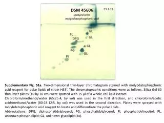
Supplementary Fig. S1a. Two-dimensional thin-layer chromatogram stained with molybdatophosphoric acid reagent for polar lipids of strain H53T. The chromatographic conditions were as follows. Silica Gel 60 thin-layer plates (10 by 10 cm) were spotted with 15 μl of a whole-cell lipid extract. Chloroform/methanol/water (65:25:4, by vol) was used in the first direction, and chloroform/acetic acid/methanol/water (80:18:12:5, by vol) was used in the second direction. Plates were sprayed with molybdatophosphoric acid reagent to locate and differentiate the polar lipids. Abbreviations: DPG, diphosphatidylglycerol; PG, phosphatidylglycerol; PI, phosphatidylinositol; PL, unknown phospholipid; GL, unknown glycolipid (4x).
Supplementary Fig. S1b. Mono-dimensional thin-layer chromatogram revealed by Dragendorff to detect phosphatidylcholine (PC) of strain H53T. PC