Download
1 / 34
351 likes | 850 Views
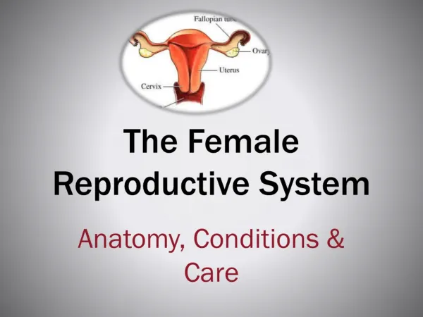
THE FEMALE REPRODUCTIVE TRACT. April 5, 2010. OBJECTIVES. Review the anatomy of the external and internal female reproductive tract with descriptive illustrations Provide some clinical correlations, i.e. physiologic changes and implications. THE EXTERNAL FEMALE GENITALIA. Vulva or pudenda.

E N D
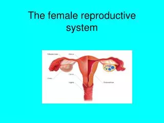
THE FEMALE REPRODUCTIVE TRACT April 5, 2010
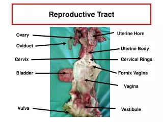
OBJECTIVES • Review the anatomy of the external and internal female reproductive tract with descriptive illustrations • Provide some clinical correlations, i.e. physiologic changes and implications
Vulva or pudenda • from the mons pubis anteriorly to the rectum posteriorly and from one lateral genitocrural fold to the other • keratinized, stratified squamousepithelium; becomes thicker, more pigmented, and more keratinized as the distance from the vagina increases
Mons Pubis (monsveneris) • fat-filled cushion; directly anterior and superior to the symphysis pubis • rounded eminence that becomes hairy after puberty; escutcheon is triangular, but may vary due to genetic and racial differences • A diamond (male) pattern can be found in one of four women
Labia Majora • two large, longitudinal, cutaneous folds of adipose and fibrous tissue. • approximately 7 to 8 cm in length and 2 to 3 cm in width; extend from the mons pubis anteriorly to become lost in the skin between the vagina and the anus in the area of the posterior fourchette
Skin: pigmented and covered with hair follicles - inner surface does not have hair follicles but has many sebaceous glands. • Usually the labia atrophy after menopause. The labia majora are homologous to the scrotum in the male.
Labia Minora(nymphae) • small, red cutaneous folds situated between the labia majora and the vaginal orifice • more delicate, shorter, and thinner than the labia majora • Divide anteriorlyat the clitoris to form the prepucesuperiorly and inferiorly the frenulum of the clitoris
Histologicallycomposed of dense connective tissue with erectile tissue and elastic fibers, rather than adipose tissue. • Skin: less cornified and has many sebaceous glands but no hair follicles or sweat glands • relatively more prominent in children and postmenopausal women • homologous to the penile urethra and part of the skin of the penis in males.
Clitoris • short, cylindrical, erectile organ at the superior portion of the vestibule • normal adult glans clitoris has a width less than 1 cm, with an average length of 1.5 to 2 cm • Size affected by previous childbearing NOT age, weight, and oral contraceptive • glans: distal one third; has many nerve endings
consists of a base of two crura, attached to the periosteum of the symphysispubis; • Body: has two cylindrical corpora cavernosa composed of thin-walled, vascular channels that function as erectile tissue • The clitoris is the female homologue of the penis in the male.
Vestibule • the lowest portion of the embryonic urogenitalsinus; the cleft between the labia minoravisualized when the labia are held apart • extends from the clitoris to the posterior fourchette • Fossanavicularis • posterior portion of the vestibule between the fourchette and the vaginal opening
Urethra • membranous conduit for urine • In females: measures 3.5 to 5 cm in length; distal orifice: 4-6mm in diameter • proximal two thirds: mucosa composed of stratified transitional epithelium • distal one third: stratified squamousepithelium and the mucosal edges grossly appear everted.
Skene's Glands (paraurethral glands) • branched, tubular glands adjacent to the distal urethra; • usually run parallel to the long axis of the urethra for approximately 1 cm before opening into the distal urethra, or may open into the area just outside the urethral orifice • the largest of the <paraurethral> glands; many smaller glands are also present • homologous to the prostate in the male.
Bartholin's Glands • vulvovaginalglands located beneath the fascia at about 4 and 8 o'clock, on the posterolateral aspect of the vaginal orifice • about the size of a pea; open into a groove between the hymen and the labia minora • Histologically composed of cuboidalepithelium, with ducts lined by transitional epithelium approximately 2 cm in length. • Homologous to Cowper's glands in the male
Vestibular Bulbs • two elongated masses of erectile tissue on either side of the vaginal orifice • immediately below the bulbocavernosusmuscle; the distal ends of the vestibular bulbs are adjacent to Bartholin'sglands • homologous to the bulb of the penis in the male
Hymen • a thin, usually perforated membrane at the entrance of the vagina • variations in the structure and shape of the hymen
histologically covered by stratified squamous epithelium on both sides, with fibrous tissue and a few small blood vessels • carunculaemyrtiformes: tags, or nodules, of firm fibrous material; remnants of the hymen identified in adult females.
Vagina • a thin-walled, distensible, fibromuscular tube that extends from the vestibule of the vulva to the uterus. • The walls of the vagina are normally in apposition and flattened in the anteroposteriordiameter; but the potential space of the vagina is larger in the middle and upper thirds. • held in position by the surrounding endopelvic fascia and ligaments
Upper portion • close to the horizontal plane when a woman is standing • supported by the upper portions of the cardinal ligaments and the parametria • Middle portion • supported by the levatorani muscles and the lower portion of the cardinal ligaments • Lower portion • in close relationship with the urogenital and pelvic diaphragms
Rugae • numerous transverse folds, prominent in the lower third, in reproductive age women; provide accordion-like distensibility • Fornices • spaces between the cervix and attachment of the vagina; the posterior fornix is considerably larger than the anterior fornix, thusa • anterior vaginal length ~6 to 9 cm • posterior vaginal length ~8 to 12 cm
Histology: • composed of four distinct layers: • Mucosa: stratified, nonkeratinizedsquamous epithelium • May become keratinized • Similar to the exocervix, but has larger and more numerous papillae • does not normally have glands • Lamina propria (tunica): fibrous connective tissue • collagen and elastic tissue, with a rich supply of vascular and lymphatic channels • The muscular layer: many interlacing fibers; with an inner circular layer and an outer longitudinal layer • Cellular areolar connective tissue: large plexus of blood vessels.
Vascular supply • Arterial supply: extensive anastomotic network throughout the vaginal length • vaginal artery: originates either directly from the uterine artery or as a branch of the internal iliac artery • may be multiple on each side of the pelvis • with an anastomosis with the cervical branch of the uterine arteries azygosarteries • Also has contributions from the internal pudendal, inferior vesical, and middle hemorrhoidal
Venous drainage: complex, accompanies the arterial system • Pudendal veins: principal drainage below the pelvic floor • Vaginal, uterine, vesical veins, as well as those around the rectosigmoid, provide drainage of the venous plexuses surrounding the middle and upper vagina
Lymphatics • characterized by wide distribution and frequent crossovers between the right and left sides of the pelvis • Upper third of the vagina: external iliac nodes • Middle third of the vagina: common and internal iliac nodes • Lower third: complex and variable distribution, including the common iliac, superficial inguinal, and perirectalnodes
Nerve supply • The nerve supply of the vagina comes from the autonomic nervous system's vaginal plexus,and sensory fibers come from the pudendal nerve. • Pain fibers: enter the spinal cord in sacral segments two to four; • With a paucity of free nerve endings in the upper two thirds of the vagina
Perineum • Pelvic diaphragm • forms the inferior border of the abdominopelviccavity; composed of a broad, funnel-shaped sling of fascia and muscle • it extends from the symphysis pubis to the coccyx and from one lateral sidewall to the other • the primary muscles of the pelvic diaphragm are the levatorani and the coccygeus
Urogenital Diaphragm • also called the triangular ligament; a strong, muscular membrane that occupies the area between the symphysis pubis and ischialtuberosities • external and inferior to the pelvic diaphragm • suspends the urethra from the pubic bone by continuations of the fascial layers • the free edge of the diaphragm is strengthened by the superficial transverse perinealmuscle • inserts posteriorly into the central point of the perineum
Perineal Body • the median raphe of the levatorani, between the anus and the vagina, reinforced by the central tendon of the perineum. • the bulbocavernosus, superficial transverse perineal, and external anal sphincter muscles also converge on the central tendon
CLINICAL CORRELATIONS • Susceptibility to infection, especially along the intertriginous areas • Changes post-menopause • Bartholin’s duct cyst – most common enlarged cyst of the vulva; abscess and urethral diverticula formation • Vulvar trauma, i.e. due to saddle injuries or childbirth and the blood supply • Continuity between the labia majora, mons pubis, and anterior abdominal wall via the subcutaneous tissue
Clinical correlations • The posterior fornix as an important surgical landmark • The distal course of the ureteras an important consideration in vaginal surgery, and the anatomic proximity and interrelationships of the vascular and lymphatic networks of the bladder and vagina • Gartner's duct cyst (cystic dilation of the embryonic mesonephros) vs. large urethral diverticula • Vaginal lubrication during intercourse and the rich vascularization of the organ • The anatomic relationship between the long axis of the vagina and other pelvic organs when altered by pelvic relaxation, i.e. from the trauma of childbirth • Atrophy or weakness of the endopelvic fascia and the development of a cystocele, rectocele, or enterocele. • rare complication : massive hemorrhage from the inferior gluteal or pudendalarteries
References • Katz et al. (2007). Comprehensive Gynecology, 5th ed. • Cunningham et al. Williams Obstetrics, 22nd edition.