Download
1 / 83
830 likes | 843 Views
Explore the biological determinants of behavior, the anatomy of the neuron, and facts about the human brain. Learn about the structure, organization, and function of the brain, neurotransmitters, and more. Discover the complex interplay of genetic, developmental, biochemical, and psychosocial factors influencing behavior.

E N D
Brain & Behaviour Abdul-Monaf Al-Jadiry, MD; FRCPsych Professor of Psychiatry
Determinants of Behaviour • Biological Determinants • Genetic Influences • Growth and developmental Influences • Biochemical Influences • Psychophysiological parameters • Learning • Psychosocial factors • Sociocultural factors
Biological Determinants of Behaviour • The complexity of the behavior of any individual is related to the complexity of its nervous system. • Generally, individuals with complex nervous systems have a greater capacity to learn new responses and thus adjust their behavior.
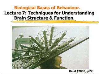
Brain & Behaviour Scientific understanding of human behaviour and experience in health and disease requires knowledge about : • Functional Anatomy of the Neuron • Functional Organization of the Brain • Neurotransmitters • Receptors • Molecular Neurobiology • Molecular Psychopharmacology
Brain & Behaviour Advances in the understanding of the structure, organization, and function of the brain offer powerful new methods for: • evaluating behaviour • diagnosing mental disorders • understanding pathophysiology of Mental Disorders • developing specific and effective therapies for Mental Disorders
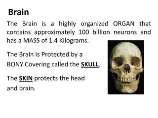
Human Brain, Some Facts • The brain is one of the largest and most complex organs in the body. • It is the upper most part of the nervous system. • The brain monitors and regulates the body's actions and reactions. • It continuously receives sensory information, and rapidly analyzes the information and then responds.
Human Brain, Some Facts • The brain is surrounded by 3 layers of tissue called the “meninges”. • The brain suspended in a fluid called “cerebrospinal fluid (C.S.F)” • The CSF is isolated from the • blood stream by the “blood-brain • barrier”. • The skull (cranium) helps protect the brain from injury.
Human Brain, Some Facts • The adult human brain weighs on average about (1.5 kg) • Men's brains are on average 100g heavier than a woman‘s • The size of the brain is around 1130 (cm3) in women and 1260 cm3 in men • The brain is made up of over 100 billion nerve Cells (Neurons) that communicate in trillions of connections called “synapses”. • At the age of 20, a man has around 176,000 km and a woman about 149,000 km of myelinated axons in their brains.
The Brain, anatomical parts The brain is made up of many specialized areas that work together:• The Cerebrum (cerebral hemispheres). • The Brain Stem, between the spinal cord and the rest of the brain. • The Cerebellum, is at the base and the back of the brain.
Functional Anatomy of the Neuron The “Neuron” • Is a cell type that is highly specialized, both anatomically and biochemically, to carry out the functions of information signaling and processing. • Hundreds of specialized types of neurons, each type subserving specialized functions. • Neurons do not divide once they are mature
Functional Anatomy of the Neuron • Neurons are composed of 4 components: • Cell body (perikaryon) • Dendrites • Axon • Presynaptic terminal
Structure of the Neuron • Cell body (Perikaryon): Consists of: • The nucleuscontains a nucleolus (plus a Barr body in females) • The cytoplasmcontains inclusions: • Nissl substance (involved in protein synthesis) • Mitochondria (involved in energy productions) • Microtubules (involved in transport of substances) • Lisosomes (bodies containing powerful enzymes) - • Golgi apparatus (involved in synthetic activities?) • Microfilaments (unknown function) • Melanin pigment ( found in substantia nigra and locus coeruleus)
Cell Nucleus The nucleus • Controls reactions in the cytoplasm by controlling the formation of proteins and enzymes. • Stores information needed for when the cell divides • Is the place where the transcription of genes and mRNA splicing occur. • The nucleus is surrounded by a double membrane, The inner and outer membrane fuse at regular spaces, forming nuclear pores • The outer membrane has ribosomes. • Ribosomes are involved in protein biosynthesis, the process of translating RNA into protein.
Cell Nucleus • The nucleus contains the chromosomes and a nucleolus. • Chromosomes are thread like strand of DNA and associated proteins that carry the genes in a linear order and function in the transmission of hereditary information. • Nucleolus is a small rounded granular body composed of protein and ribonucleic (RNA) and involved in ribosomal RNA synthesis and the formation of ribosomes |
Structure of the Neuron 2.The Axon • Usually single • The proximal portion is called the “Axon Hillock” • Branches distally - each branch forms an outpouch at its end called the “Button” • Myelinated and unmyelinated • Conducts impulses away from the perikaryon
Structure of the Neuron 3. Dendrites • Usually more than one per neuron • Contain Nissl substance • Branched are studded with dendritic spines (sites for synaptic contact) • Conduct information to the perikaryon
The Synapse • Is a specialized structure involved in the transmission of information from one neuron to another • The “Synapse consists of: • Button: outpouch of the terminal portion of the axon of the Presynaptic neuron • Dendritic membrane of the adjacent postsynaptic neuron(dendritic spine) • Neurotransmission is accomplished by: • Chemical Transmission by messengers called “Neurotransmitters (NTs)” • Electrical Transmission by nerve impulses and action potentials
Receptors • The dendritic membrane at the synapse is markedly enriched with “Receptors” that respond to the neurotransmitter released by the terminal button of the Presynaptic neuron. • Neuroreceptors are proteins that span the neuronal membrane. • Receptors have: - ligand binding regions- that are accessible to extracellular messengers - ligand-gated channels consist of channel pores
Developmental Brain OrganizationBrain structures as derivatives of the neural tube Primary vesiclessecondary vesiclesBrain components - Prosencephalon Telencephalon Cerebral Cortex (forebrain) Hippocampus Amygdala Striatum Diencephalon Thalamus & subthalamus Hypothalamus, Epithalamus - Mesencephalon Mesencephalon Midbrain (midbrain) - Rhombencephalon Metencephalon Pons (hindbrain) Cerebellum Myelencephalon Medulla
Functional Brain Systems Three functional brain systems illustrate the relation between the organizational principles and the structural components of the human brain: • Thalamocortical system • Basal ganglia system • Limbic System
1. Thalamocortical system • The connection between the thalamus, the cortex, and certain related structures • Comprises 3 thalamocortical systems (each with different pattern of functional circuity): • Sensory System, • Motor System, • Association System
2. Basal Ganglia System • A collection of nuclei grouped together on the basis of their interconnections • Play an important role in: • regulating movement • cognitive functions
Basal Ganglia System • Major components: 1. Caudate 2. Lentiform nucleus = putamen + Globus pallidus (pallidum or paleo striatum) 3. Subthalamic nucleus 4. Substantia nigra [Striatum = all the above nuclei]
Thank you Thank You
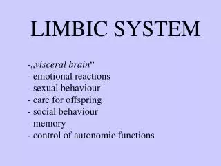
Psychophysiological Determinants of Behaviour • The Limbic System • Reticular activating System (ARAS) • Cortical Sites (Cerebral Cortex)
The Limbic System • The limbic system is a set of brain structures that forms the inner border of the cerebral cortex. • The structures include the hippocampusand amygdala that support a variety of functions including emotion, behavior and long term memory. • The word “Limbic” = Latin word “Limbus” (border) applied by “Pierre Broca” (French physician, surgeon, anatomist and anthropologist) more than 100 years ago.
Limbic System • The term Limbic system applied by “Paul MacLean, 1952” to describe the circuity that relates certain telencephalic (Forebrain) structures and their connections with the hypothalamus and its output pathway that control autonomic, somatic, and endocrine functions.
limbic lobe • The limbic lobe is named by Broca in1878. • He identified it with the Cingulate and Parahippocampal gyri, and associated it with the sense of smell. • The limbic lobe is an arc-shaped region of cortex on the medial surface of each cerebral hemisphere, consisting of parts of the frontal, parietal and temporal lobes. • A direct connection between emotion and the limbic lobe has been identified.
Limbic Structures Amygdala: • A Latin word means 'almond', 'tonsil‘. • Are almond-shaped groups of nuclei located deep within the medial temporal lobes of the brain. • Has a primary role in the processing of memory and emotional reactions • Involved in signaling the cortex of motivationally significant stimuli such as those related to reward and fear in addition to social functions such as mating.
Hippocampus • Is a Latin word meaning “seahorse“ or "sea monster” • Aranzi (1587) likened it to a seahorse, or alternatively to a silkworm. • It is the ridge running along the floor of the temporal horn of the lateral ventricle in the medial temporal lobe underneath the cortical surface. • Plays important roles in the consolidation of information from short-term memory to long-term memory. • It contains two main interlocking parts: Ammon's hornand the dentate gyrus.
Hippocampus • The hippocampus is one of the first regions of the brain to suffer damage in Alzheimer's disease. • Extensive, bilateral hippocampal damage may cause anterograde amnesia. • Many reports have found reductions in the size of the hippocampus in schizophrenic subjects
Parahippocampal gyrus • Parahippocampal gyrus: surrounds the hippocampus. • It plays an important role in memory encoding and retrieval. • Plays a role in the formation of spatial memory. • Parahippocampal Asymmetry has been observed in schizophrenia.
Cingulate gyrus • The Cingulate gyrusis situated in the medial aspect of the cerebral hemisphere, lies immediately above the corpus callosum. • It is involved with emotion, learning, memory, executive function and respiratory control • The Cingulate cortex is highly important in disorders such as depression and schizophrenia. • Anterior cingulate gyrus was found to be smaller in schizophrenic patients.
Hypothalamus • The hypothalamus (from Greekunder and room, chamber). • Contains a number of small nuclei with a variety of functions. • Located below the thalamus, just above the brain stem. • It is roughly the size of an almond. • One of the most important functions of the hypothalamus is to link the nervous system to the endocrine system via the pituitary gland (hypophysis).
Hypothalamus • The hypothalamus is responsible for certain metabolic processes and other activities of the autonomic nervous system. • It synthesizes certain neurohormones, often called hypothalamic-releasing hormones, and these in turn stimulate or inhibit the secretion of pituitaryhormones. • The hypothalamus controls body temperature, hunger, thirst, fatigue, sleep, and circadian cycles.
Limbic Structures Fornix: carries signals from hippocampus to the mammillary bodies and septal nuclei. Mammillary bodies: Important for the formation of memory. Septal nuclei: Located anterior to the interventricular septum, they provide critical interconnections Parahippocampal gyrus: Plays a role in the formation of spatial memory Dentate gyrus: contributes to new memories
Thalamus • The thalamus (from Greekθάλαμος, "inner chamber) • It is a midline symmetrical structure within the brain, situated between the cerebral cortex and midbrain and surrounds the 3rd ventricle. • Its function includes: • relaying sensory and motorsignalsto the cerebral cortex, • regulation of consciousness, sleep, and alertness.