Download
1 / 32
330 likes | 784 Views
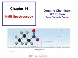
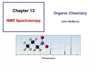
Chapter 13 NMR Spectroscopy. NMR - Nuclear Magnetic Resonance NMR is a form of spectroscopy that uses an instrument with a powerful magnet to analyze organic compounds. Invented by physicists (1950’s), then used by chemists (1960’s). Why is it called NMR?. Nuclear Magnetic Resonance

E N D
Chapter 13 NMR Spectroscopy NMR - Nuclear Magnetic Resonance NMR is a form of spectroscopy that uses an instrument with a powerful magnet to analyze organic compounds. Invented by physicists (1950’s), then used by chemists (1960’s).
Why is it called NMR? Nuclear Magnetic Resonance Nuclear – because it looks at the nucleus of an atom, most commonly a hydrogen atom. A hydrogen atom nucleus consists of one proton with a +1 charge and “spin” of ½. It acts like a tiny bar magnet. generatesmagneticfield
NMR – Effect of Magnetic Field No external magnetic field applied to sample Random orientation of nuclear spins Sample placed in an external magnetic field
NMR: Absorption of Energy Initial State – nucleus at low energy level
NMR: Information Obtained from a Spectrum • An NMR Spectrum will generally provide three types of information: • Chemical Shift – indicates the electronic environment of the nucleus (shielded or deshielded) • Integration – gives the relative number of nuclei producing a given signal • Spin-Spin Coupling – describes the connectivity
1H NMR Spectrum – H2O A sample of water is placed in an NMR instrument, and a proton spectrum is recorded (scanned from left to right).
When does nucleus absorb energy? 3. 2, External Field (Ho)from magnet
NMR: Simple 1H NMR Spectrum Showing Chemical Shift Two types of protons (a CH2 and a CH3) give two separate signals at two different chemical shifts.
NMR: Chemical Shift Practice EN Group -O-CH3 -Si-CH3 -C-CH3 Cl3C-H Assign the four groups shown to the four NMR singals, based on each element’s electronegativity.
NMR: Chemical Shift Regions Chemical shift zero is set to TMS (tetramethylsilane), a reference compound Chemical shift measured in ppm.
NMR: Chemical Shift Regions Alkane Region (high electron density): Heteroatom Region: Double Bond Region:
NMR: Chemical Equivalence and Number of Signals How many signals will the following compounds show in their 1H NMR Spectrum? (Hint: check for symmetry)
NMR: Chemical Equivalence and Number of Signals How many signals should appear in the proton NMR spectrum for these compounds? In theory: Signals actually resolved:
NMR: Overlapping Proton Signals Protons b, c, and d are all nearly the same, and their signals are not resolved in this spectrum.
Review: How Many NMR Signals? How many signals will the following compounds show in their 1H NMR Spectrum? (Hint: check for symmetry) No rotation about double bonds
NMR: Chloroethane Fast rotation around single bonds gives an “averaged” spectrum for the three methyl hydrogens.
NMR: A Second Proton Spectrum Note: the signal for the nine methyl H’s is larger than the CH2 signal
NMR: Integration Indicates Relative Number of Nuclei The height of the integration line (“integral”) gives you the relative number of nuclei producing each signal.
NMR: Splitting into a Doublet doublet Note that the signal at 1.6 ppm for the methyl group is split into two peaks. Remember that this is one signal, composed of two separate peaks.
NMR: Signal Splitting, n+1 Rule • A signal is often split into multiple peaks due to interactions with protons on carbons next door. Called spin-spin splitting • The splitting is into one more peak than the number of H’s on adjacent carbons (“n+1 rule”) • Splitting of a signal can give doublets (two peaks), triplets (three peaks), quartets (4 peaks), ect. • The relative intensities given by Pascal’s Triangle: doublet 1 : 1 triplet 1 : 2 : 1 quartet 1 : 3 : 3 : 1 pentet: 1 : 4 : 6 : 4 : 1
NMR: Signal Splitting, n+1 Rule n+1 Rule: A signal in the proton NMR spectrum will be split into n+1 peaks, where n is the number of protons on adjacent carbons. Example: CH3-CH2-Br For the Methyl Group – There are two protons ‘next door’ (n=2), so the methyl signal will be split into three peaks (2+1), which is called a triplet. For the -CH2- Group: Three protons next door means the CH2 signal will be split into 4 (3+1) peaks, called a quartet.
1H NMR Spectrum for Bromoethane integration: 2 H 3 H Note the expansions printed above
NMR: Origin of Spin-Spin Splitting Net result:
NMR: Doublets and Triplets Triplet: for the two protons next door,there are four combinations possible: αααβ βββ α Doublet: the one proton next doorcan be either up or down (α or β)
NMR: Using the n+1 Rule Using the n+1 rule, predict the 1H NMR spectrum of 2-iodopropane.Give splitting pattern, integration, and approximate chemical shift. Note that the methyl groups are equivalent, so they will give one signal in the NMR spectrum.
doublet Seven line pattern NMR: Spectrum of 2-iodopropane
NMR: Rules for Spin-Spin Splitting • The signal of a proton with n equivalent neighboring H’s is split into n + 1 peaks • Protons farther than two carbon atoms apart do not split each other • Equivalent protons do not split each other
Common 1H NMR Patterns 1. triplet (3H) + quartet (2H) 2. doublet (1H) + doublet (1H) 3. large singlet (9H) 4. singlet 3.5 ppm (3H) 5. large double (6H) + muliplet (1H) 6. singlet 2.1 ppm (3H)
Common 1H NMR Patterns 7. multiplet ~7.2 ppm (5H) 8. multiplet ~7.2 ppm (4H) 9. broad singlet, variable chemical shift