Download
1 / 16
160 likes | 187 Views
Designing a technique to measure capillary recruitment in mice for studying MGU and microcirculation, crucial for understanding metabolic processes like diabetes. Using methods like confocal microscopy to capture “honeycomb” structures. Presenting past techniques and future plans for further research.

E N D
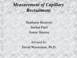
Stephanie Beatrous Snehal Patel Samar Sharma Advised by: David Wasserman, Ph.D. Measurement of Capillary Recruitment
Project Objective • To design a technique to measure capillary recruitment in vivo or in situ in the skeletal muscle microvasculature of mice for the purpose of investigating basal and insulin-stimulated MGU.
Regulation of Microcirculation QM= Blood Flow to Muscle QC= Blood Flow per Capillary NC= Number of Perfused Capillaries VC= Blood Velocity AC= Capillary Cross Sectional Area QM=QCNC=VCACNC • Blood flow to the muscles is directly proportional to the number of perfused capillaries (NC) • Surface area for glucose exchange is proportional to NC, so a higher blood velocity will not be as efficient
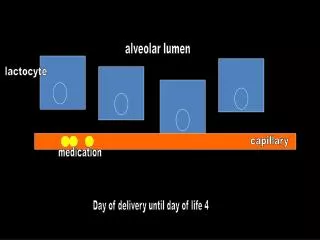
Capillary Recruitment • An increase in the number of perfused capillaries is known as capillary recruitment • 10% of capillaries are perfused in resting skeletal muscles • Capillary density is a function of metabolic requirements
Muscle Glucose Uptake • Muscle Glucose Uptake (MGU) is a linear sequence of three processes. • Glucose delivery from blood to the interstitial space • Glucose transport from interstitial space to the intercellular environment. • Glucose phosphorylation by Hexokinase II
Significance of MGU • Glucose plays a role in cell function by serving as an energy source. • Each of the three processes in MGU determine whether cellular needs for energy source are met. • As MGU is controlled by many physiological subsystems, alterations could lead to pathology.
Diabetes • Diabetes is fundamentally a disorder of MGU. • 16million Americans • 98billion dollars in costs • 18% of deaths (>25yrs old). • 7th leading cause of death in 1993 • pharminfo.com
Methods • 1.1 ug/mL saline solution of TX Red-Dextran T70 infused at 1.375 uL/min • Dye solution equilibrated in mouse after 100 min • Catheterized mouse anesthetized using Nambitol • Surgery performed, vasus lateralus muscle exposed on right leg • Mouse mounted on confocal microscope • 50 uL sodium nitroproside (SNP) injected to vasodilate • Observations made and images collected
Results • “Honeycomb” structure imaged • Capillary flow evident in structure
Past Work • Research of current imaging techniques • Optical Coherence Tomography (OCT) • Intravital Microscopy • Microspheres • Functional Magnetic Resonance Imaging (fMRI) • Fluorescent Microscopy • Two-photon Excitation Microscopy • Whole-Field Microscopy • Widefield Imaging • Confocal Microscopy
Current Status • Research almost complete • Need to continue assessing feasibility of methods • Most Promising method identified • Fluorescent Microscopy
Future Plans • Test Fluorescent Microscopy on 2/16/01 using the confocal microscope and implemented technique. • Adapt this technique to study MGU. • If adaptation unsuccessful, use Optical Coherence Tomography (OCT)
Future Plans (cont.) • Test variations in the technique • Refine protocol for measuring capillary recruitment • Obtain images and assess quality of results