Download
1 / 46
540 likes | 1.44k Views
Myofascial pain dysfunction syndrome. TMJ Dysfunction Syndrome/ Myofascial Pain Dysfunction Syndrome. History- Costen (1934)- occlusal etiology in TMJ pain.

E N D
TMJ Dysfunction Syndrome/ MyofascialPain Dysfunction Syndrome History- • Costen (1934)- occlusal etiology in TMJ pain. • He reported association of bite like ear pain, sinus pain, decreased hearing, tinnitus, dizziness, burning & vertigo & occipital headaches. • Scwartz (1956)- TMJ pain dysfunction syndrome & blamed the masticatory & perimasticatory musculature leading towards the symptoms. • He noted altered psychologic make up and advocated use of muscle relaxants, restrictions of oral openings for resting the muscles. • Neelima Malik, Textbook Of Oral & Maxillofacial Surgery, 2nd Edi
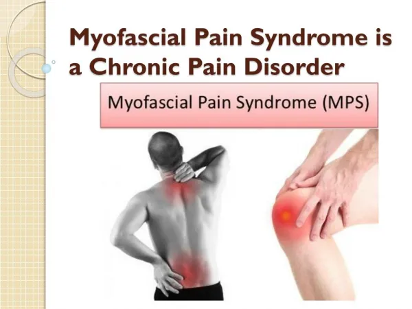
Laskin (1969)- Myofascial Pain Dysfunction Syndrome. • He implicated psychophysiologic theory stating that psychological stress leads to myospasm & advised tranquilizers, muscle relaxants. Definition • MPDS is a pain disorder, in which unilateral pain is referred from the trigger points in myofascial structures, to the muscles of the head and neck. • MPDS is the most common cause of masticatory pain & limited function for which patient seeks dental consultation & the source of the pain treatment.
Etiology • Tooth muscle theory- occlusal interference cause an allteredproprioceptive feedback, leading to incoordination & spasm of muscles of mastication. • Prosthetic problems- fauty prosthesis, over closure, bilateral loss of molar teeth, increased vertical dimentions. • Malocclusion- by Oral habits (clenching, grinding of teeth), anxiety. • Psychophysiologic theory- masticatory muscle spasm causes MPDS, degenerative arthritis & contracture arthritis. • Steep angulation of articular eminence. Okeson, Jeffrey P. (2003). Textbook of Management of temporomandibular disorders and occlusion (5th ed.)
Okeson, Jeffrey P. (2003). Textbook of Management of temporomandibular disorders and occlusion (5th ed.)
Clinical characteristics Laskin’s Cardinal symptoms of MPDS :- • Pain or discomfort anywhere about the head or neck. • Limitation of motion of the jaw. • Joint noises– grating, clicking, snapping. • Tenderness on palpation of the muscles of mastication. Negative characteristics:- • Absence of clinical, radiographic or biochemical evidence of organic changes in TMJ. • Lack of tenderness in TMJ area when palpated via external auditory meatus. • A zone of reference • Trigger points in muscles • Occasional associated symptoms • The presence of contributing factors
Zone of reference • Referral of myofascial trigger point pain of temporalis muscles refers only to the maxillary teeth.
The masseter refers only to the posterior teeth. • The digastric anterior refers only to the mandibular incisors.
Trigger points • Trigger points exist as a localized tender areas within taut bands of skeletal muscles & stimulated by macro & microtraumatic episodes, they refer a characteristic pain pattern to a distant group of muscles, i.e. zone of reference. • Palpation of trigger points gives a positive ‘jump sign’.
Other criteria • Psychologic or central etiology- • Muscle fatigue by unusual habits & occlusal disharmony. • Occlusal or peripheral etiology- • Inherent malocclusion- developmental deformities • Acquired malocclusion • Iatrogenic • Intrinsic joint disorders • Neelima Malik, Textbook Of Oral & Maxillofacial Surgery, 2nd Edi
History of the patient • Mode of onset, duration, frequency & quality of pain. Site & reference point of pain. • Time of the day, at which pain is most pronounced. • Occupation. • Sleeping habits. • Parafunctional habits. • H/O previous trauma, prolonged dental work etc. • Family or emotional problems. • Associated symptoms. • Aggravating & relieving factors. • Neelima Malik, Textbook Of Oral & Maxillofacial Surgery, 2nd Edi
Physical examination • Articular / joint examination • Dental examination • Muscular examination • Cervical examination • Neelima Malik, Textbook Of Oral & Maxillofacial Surgery, 2nd Edi
Clinical examination of TMJ • TMJ pain- • Determined by digital palpation of the joints – when mandible is in both stationary as well as dynamic movement • Finger tips placed – lateral aspect of both the joints – feel for the lateral poles passing downwards & forwards across articular eminence on repeated opening & closing. • A medial force is then applied to – record symptoms if any in static position & then on opening & closing. • Also, on maximal opening – fingers are rotated slightly posteriorly to apply force on the posterior aspect of the condyle (to evaluate posterior capsulitis & retrodiscitis)
Temporalis muscle • Palpation of the posterior, middle, and anterior regions and tendon of the Temporalis. • I / o- The finger is moved up the anterior border of the ramus until the coronoid process and the attachment of the tendon of the temporalis are felt.
Massater muscle • Palpation of the masseter muscles at their superior attachment to the zygomatic arches. • Palpation of the superficial masseter muscles near the lower border of the mandible.
Functional manipulation of inferior lateral pterygoid. • The patient is asked to protrude against resistance provided by the examiner. • Functional manipulation of the superior lateral Pterygoid is achieved by asking the patient to bite on at tongue blade bilaterally.
Medial pterygoid • Tell patient to open mouth wide, protrude against resistance, clench the teeth together, and then bite on a separator when these function causes pain then medial pterygoid muscle is involved.
Sternocleidomastoid • Palpation of the sternocleidomastoid muscle high near the mastoid process&low near the clavicle.
Articular or TMJ Function and Range of Motion:- 1. Amount of oral opening and the excursions. 2. Extent of movement i) ROM – Range of motion i) AROM – Active range of motion iii) PROM – Passive range of motion • Palpation for tenderness. • Grading of click or crepitation- noises evaluation. • Auscultation (stethoscopic evaluation), if needed. • Neelima Malik, Textbook Of Oral & Maxillofacial Surgery, 2nd Edi
1. Amount of oral opening and excursions. • Opening path & amount of deviation should be noted. • Early opening deviation is due to spasm lateral pterygoid muscle of same side. • Normal range of protrusive movement- 10mm • Lateral excursions- normal- 10mm • Pain or inflammation indicates one of the following conditions • Joint inflammation, • muscle dysfunction. • Anteriorly displaced disc etc • Neelima Malik, Textbook Of Oral & Maxillofacial Surgery, 2nd Edi
2. Extent of movement • ROM – Range of motion- • Normal vertical ROM in adults is-40- 50 mm • Hypomobility without pain gives indication for pathology. • Measurement of maximum pain free motion is noted. • Limited AROM with pain indicates structural restrictions by muscular problems. • PROM tests all inert structures. • Neelima Malik, Textbook Of Oral & Maxillofacial Surgery, 2nd Edi
3. Areas of tenderness on palpation • Simultaneous palpation of both the joints with index fingers laterally over the joints & through the external auditory canal in open & closed position. • Pain is unrelated to closure in posterior joint palpation may indicate ear problem or inflammation. • Neelima Malik, Textbook Of Oral & Maxillofacial Surgery, 2nd Edi
4. Timing of the click • Noted whether it is coming during opening, closing or both should be ascertained. • Distinct sound click • Crepitus • Multiple scraping, grating noises These sound can be heard by stethoscope. • Neelima Malik, Textbook Of Oral & Maxillofacial Surgery, 2nd Edi
Recent diagnostic methods:- • TMJ arthrography. • Computed radiography (CR). • Computed Tomography (CT) scan & Magnetic Resonance Imaging (MRI). • Bone Scintigraphy- nuclear imaging • Single Photon Emission Computerized Tomography (SPECT). • Neelima Malik, Textbook Of Oral & Maxillofacial Surgery, 2nd Edi
Phase I therapy psychophysiologic discussion. Home therapy (diet & exercise) Muscle relaxant & NSAIDs (2-4 wks)- 50% resolution Laskin and Block, (1986) Phase Il therapy Home therapy and medications + bite appliance (2 to 4 wks) 20% to 25%resolution. Phase lll Physiotherapy (ultrasound, electrogalvanic stimulation) or relaxation therapy (yoga, biofeedback) (4 to 6 weeks) 10% to 15% resolution. Phase lV Psychologiccounselling pain clinic
Medications • NSAIDs to reduce inflammation & pain in muscles & joint. • Aspirin : 2 tabs 0.3 to 0.6gm/ 4 hourly.(ECOSPIRIN) • Piroxicam: cap. 10 to 20mg /once daily.(FELDENE) • Ibuprofen : 200 to 600mg/3 times a day.(BRUFEN) • Pentazocine: 30 mg i.v./i.m./s.c. every 3– 4 hrs max.- 360mg. (TALWIN, TALACEN) • Muscle relaxant- • Methocarbamol : muscle relaxant- 1500mg/ 4 times a day for 2-3 days,1000mg i.v./ 8 hrly (ROBAXIN) • Metaxalone- (SKELAXIN) • Chlorzoxazone - (FLEXON MR)- 400 mg, 325mg, 250 mg • Antidepressant- • Diazepam- (VALIUM, CALMPOSE) & chlordiazepoxide (sedative) 5 to 10mg /2 to 3 times a day.(LIBRIUM) • Amitriptyline: - 25mg/ 3- 4 times a day or at bedtime.(ELAVIL, VANATRIP). • Tripathi, textbook of pharmacology, 5th edi., • NeelimaMalik, Textbook Of Oral & Maxillofacial Surgery, 2nd Edi
Trigger Point Injection • A trigger point is located, trapped between the fingers, and injected (with a short 27-gauge needle). • 0.5% bupivacaine with epinephrine. • Blocks are given at 48-hour to weekly intervals into nerve distributions and particularly into zones of muscular trigger foci. Okeson, Jeffrey P. (2003). Textbook of Management of temporomandibular disorders and occlusion (5th ed.)
Therapeutic Anesthetic Block • The auriculotemporal nerve can be blocked by passing a 27-gauge needle through the skin just anterior to and slightly above the junction of the tragus and the earlobe. • The needle is then advanced until it touches the posterior neck of the condyle. The needle is then repositioned in a more posterior direction behind the posterior neck of the condyle. • Once the neck of the condyle is felt, the tip of the needle is carefully moved slightly behind posterior aspect of the condyle in an anteromedial direction to a depth of 1 cm. Okeson, Jeffrey P. (2003). Textbook of Management of temporomandibular disorders and occlusion (5th ed.)
Physiotherapeutic modalities • Heat application. • Ultrasound • Heat lamp • Diathermy. • Cryotherapy. • Ice pack • Vapo- coolant spray • Ice massage • Cold whirlpool • Massage with counter-irritants & vibrators. • Electro Galvanic Stimulation. • Transcutaneous Electronic Nerve Stimulator (TENS). • Active stretch exercises. Mujakperuo HR, Watson M, (2010). "Pharmacological interventions for pain in patients with temporomandibular disorders". The Cochrane Database of Systematic Reviews
Pathophysiologic effects of topical modalities Scott F. Nadler, DO, FACSM, Kurt Weingand, and Roger J. Kruse, MD, The Physiologic Basis and Clinical Applications of Cryotherapy and Thermotherapy for the Pain Practitioner Pain Physician. 2004;7:395-399.
Heat application/ thermotherapy • Thermotherapy is the therapeutic application of any substance to the body that adds heat to the body resulting in increased tissue temperature. • Heat therapy, which can be either superficial or deep, is like cryotherapy in that it provides analgesia and decreased muscle tonicity. • Unlike cryotherapy, thermotherapy increases tissue temperature, blood flow, metabolism, and connective tissue extensibility. • Heat therapy is delivered by three modes: • Ultrasound • Heat lamp • Diathermy. Scott F. Nadler, DO, FACSM, Kurt Weingand, and Roger J. Kruse, MD, The Physiologic Basis and Clinical Applications of Cryotherapy and Thermotherapy for the Pain Practitioner Pain Physician. 2004;7:395-399.
Ultrasound therapy • Ultrasound (US) is used for heating deep tissues. • It is a noninvasive method which consists of piezoelectric crystals that convert the electrical energy to mechanical oscillation energy using high-frequency alternating current (van derWindt, 1999). • 2- 3 MHz – upto 3 cm deeper tissues.
Promotes tissue repair • Contineous or pulsed • Both produces thermal effects • Contineous produces more thermal energy. • A unidirectional movement of the ultrasound field causing a micro massage of the target tissues that increases cell diffusion which is thought to promote tissue repair. Scott F. Nadler, DO, FACSM, Kurt Weingand, and Roger J. Kruse, MD, The Physiologic Basis and Clinical Applications of Cryotherapy and Thermotherapy for the Pain Practitioner Pain Physician. 2004;7:395-399.
Pulse ratio • Concentration of energy on time basis • Time of machine at on : Time of machine at off Scott F. Nadler, DO, FACSM, Kurt Weingand, and Roger J. Kruse, MD, The Physiologic Basis and Clinical Applications of Cryotherapy and Thermotherapy for the Pain Practitioner Pain Physician. 2004;7:395-399.
Phonophoresis • The use of ultrasound to enhance the delivery of topically applied medications • Hydrocortisone- 10% - wycort 2.5% ointment- TDS • Ketoprofen- 10% • Dexamethazone • Clobetasol ointment 0.05% in orabase • NSAIDs - diclofenac sodium gel as a coupling media (Álvarez-Soriaet al., 2008). • 1 MHz frequency with transducer having an affective radiating Intensity of 1.5 W/ cm² in continuous mode to insure reaching the deep tissues (Kitchen and Bazin, 2002).
Cryotherapy • Cryotherapy is defined as the therapeutic application of any substance to the body that removes heat from the body, resulting in decreased tissue temperature. • Ice pack • Ice massage • Vapo-coolant spray • Cryotherapy decreases tissue blood flow by causing vasoconstriction, and reduces tissue metabolism, oxygen utilization, inflammation, and muscle spasm. • Topical cold treatment decreases the temperature of tissues to a depth of 2 to 4 cm, decreasing the activation threshold of tissue nociceptors and the conduction velocity of pain nerve signals causing local anesthetic effect called cold-induced neuropraxia. Scott F. Nadler, DO, FACSM, The Physiologic Basis and Clinical Applications of Cryotherapy and Thermotherapy for the Pain Practitioner Pain Physician. 2004;7:395-399.
Cold application • Ice pack or direct ice massage encourages the relaxation of muscles that are in spasm and thus relieves the associated pain. During warming there is an increase in blood flow to the tissues that assists tissue repair. • Continued icing will result in a mild aching and then numbness. After a period of warming a second application may be desirable. Okeson, Jeffrey P. (2003). Textbook of Management of temporomandibular disorders and occlusion (5th ed.)
Use of vapocoolent spray • Vapocoolant sprays do not penetrate tissue as does ice, and therefore it is likely that the reduction in pain is more associated with the stimulation of cutaneous nerve fibers that in turn shut down the smaller pain fibers (the C fibers). • Ethyl chloride and fluoromethane. • Applied from a distance of 1 or 2 feet for 5 sec. • After the tissue has been rewarmed, the procedure can be repeated. • A towel can be used to protect eyes, ears, nose, mouth. • When myofascial (trigger point) pain is present, a technique described as “spray and stretch” is used. Okeson, Jeffrey P. (2003). Textbook of Management of temporomandibular disorders and occlusion (5th ed.)
Electrogalvanic stimulation therapy • Uses the principle that electrical stimulation cause muscle contraction. • EGS uses a high-voltage, low-amperage, monophasic current of varied frequency. • A rhythmic electrical impulse is applied to the muscle, creating repeated involuntary contractions and relaxations. • The intensity and frequency of these can be varied according to the desired effect, and they may help to break up myospasms, as well as increase blood flow to the muscles and leads to a reduction of pain in compromised muscle tissues. Okeson, Jeffrey P. (2003). Textbook of Management of temporomandibular disorders and occlusion (5th ed.)
Transcutaneous Electrical Nerve Stimulation. • TENS produces a continuous stimulation of cutaneous nerve fibers at a subpainful level. • This uses a low-voltage, low-amperage, biphasic current of varied frequency and is designed primarily for sensory counterstimulation in painful disorders. • When a TENS unit is placed over the tissues of a painful area, the electrical activity decreases pain perception. • When the intensity of a TENS unit is increased to the point that motor fibers are activated, this becomes an EGS unit that is no longer used for pain control but instead for muscle relaxation. Okeson, Jeffrey P. (2003). Textbook of Management of temporomandibular disorders and occlusion (5th ed.)
During TENS, pulsed currents are generated by a portable pulse generator and delivered across the intact surface of the skin via conducting pads called electrodes. • The conventional way of administering TENS is to use electrical characteristics that selectively activate large diameter ‘touch’ fibres (Aβ) without activating smaller diameter nociceptive fibres (Aδ and C). • Evidence suggests that this will produce pain relief in a similar way to ‘rubbing the pain better’. Okeson, Jeffrey P. (2003). Textbook of Management of temporomandibular disorders and occlusion (5th ed.)
Acupuncture • Acupuncture involves stimulation of the body at certain points. During a treatment, thin steel needles are inserted into the skin, then manipulated gently by hand or with light electrical stimulation. • Treatment is short (6 week) • Acupuncture needles stimulate the nervous system by releasing natural painkillers such as endorphins and serotonin. • Each patient responds to acupuncture differently. Some people notice an immediate improvement, while some need several treatments to experience the full effect. • The British Dental Acupuncture Association reported that about 70% of patients show some benefit. Gustav O Kruger ; Textbook of Oral and Maxillofacial Surgery, ch 26, 6th edi; 700-756
Biofeedback Training • The patient is encouraged to assume a relaxed position in a comfortable, quiet setting. • The electromyographic sensors are attached to the masseter muscle. • A finger sensor may also be used to monitor temperature and/or galvanic skin response. • The patient is instructed to relax the muscles as much as possible. • The computer monitor provides immediate feedback regarding the success in reducing the muscle activity. • After several training sessions the patient becomes aware of effective relaxation and is encouraged to accomplish this without the biofeedback unit. Okeson, Jeffrey P. (2003). Textbook of Management of temporomandibular disorders and occlusion (5th ed.)
Psychological approaches Gustav O Kruger ; Textbook of Oral and Maxillofacial Surgery, ch 26, 6th edi; 700-756 • Significant control over pain and other sensory complains may be gained through psychophysiological techniques such as yoga, meditation, biofeedback, hypnosis, and psychotherapy. • Hypnosis is another paraphysiological technique with primary action on pain tolerance thresholds for the control of chronic facial pain. Carefully selected patients may, with training in autosuggestion, come to effectively deny or accept their pain. • Counselling and psychotherapy should be included in the overall treatment.
![[PDF DOWNLOAD] Travell, Simons Simons' Myofascial Pain and Dysfunction: The](https://cdn5.slideserve.com/10147649/welcome-to-my-slide-dt.jpg)