Download
1 / 19
190 likes | 209 Views
Small angle X-ray scattering (SAX) Kinetic crystallography – slow reactions Kinetic crystallography – Laue method Kinetic crystallography – freezing techniques Anomalous dispersion. Small angle X-ray scattering (SAX):

E N D
Small angle X-ray scattering (SAX) Kinetic crystallography – slow reactions Kinetic crystallography – Laue method Kinetic crystallography – freezing techniques Anomalous dispersion
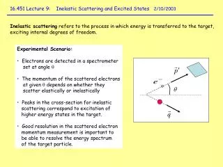
Small angle X-ray scattering (SAX): Waves are scattered at objects with which they interfere. The finer the lattice (i.e. small objects), the larger the scattering angle. X-rays (electromagnetic waves) are scattered at objects of molecular size (nm) with variing electron density. direct beam X-ray detected signal
Small angle scattering: MurA (UDP-N-acetylglucosamine enolpyruvyltransferase) The enzyme has two domains, which are in a closed conformation (with substrate and inhibitor) or an open conformation (without ligand) – in the crystal structures. Are the differences an artfact of the crystal structure or of relevance for the solution structure?
Small angle scattering: MurA open modelled closed closed
Small angle scattering: MurA Complex with pyruvate-P (•) • Fitted with open structure • Fitted with closed model • Fitted with closed structure
Small angle scattering: MurA Protein solution without and with UDP-glucosamine Fitted with open structure Fitted with closed structure -substrate fit open +substrate fit closed
Kinetic crystallography – slow reactions Remember: L-Haloacid dehalogenase. Trapping of the covalent intermediate by cryocooling (‚freezing‘) during the reaction. Data collection needs to be faster than preparation / soaking / reaction. Data collection: < 10 min at a strong X-ray source for data set, s for single frames, ns - ms for Laue frames. Preparation: s, Soaking: 10-100 s, Reaction: ?
Kinetic crystallography – Laue method Normally only single wavelength X-ray light is used for diffraction experiments: ca. 100 frames / data set. With white X-rays (l = 0.8-2 A) only few frames are needed and the intensity is higher: ms / frame.
Kinetic crystallography – Laue method Photoactive yellow protein: Light triggers a conformational change in the protein. After a short laser puls (ns), a Laue photograph (ns) is taken. The protein relaxes and the procedure is repeated.
Kinetic crystallography – Laue method Photoactive yellow protein: The photocycle
Kinetic crystallography – Laue method Photoactive yellow protein: pG groundstate chromophore H-bonded to Tyr42 trans double bond,
Kinetic crystallography – Laue method Photoactive yellow protein: pR = first excited state at 1- 1.2 ns after excitation chromophore H-bonded to Tyr42 cis double bond,
Kinetic crystallography – Laue method Photoactive yellow protein: pB = second excited state at 2-12 ms after excitation chromophore H-bonded to Arg52, cis double bond
Kinetic crystallography – Laue method Photoactive yellow protein: The photocycle pG pR pB
Kinetic crystallography – freezing techniques A crystal of a protein-substrate complex is cryocooled. The reaction is started, e.g. by laser. The reaction cannot proceed, because motions are frozen. At increasing temperature further steps may be enabled.
Kinetic crystallography – freezing techniques Myoglogin: A crystal with a CO complexed to heme is irradiated with a laser. CO dissociates from the heme. But: due to the low temperature, the CO cannot diffuse out of the binding pocket. At higher temperature the CO can be seen on its way out.
Anomalous dispersion: Normally the incident X-ray continues without phaseshift after the scatterer. If the energy is at or above the absorption edge (energy to eject an electron from the atom) of the scatterer, a phaseshift occurs: anomalous scattering. The anomalous scattering is quantified by f‘‘. It is strongly wavelength dependent.
Anomalous dispersion: Zn Cu E Use this energy to see an effect from Cu and Zn Use this energy to see an effect only from Cu Only normal scattering
Anomalous dispersion: Zn Zn Cu Cu ‚Anomalous electron density‘ calculated from anomalous scattering at Cu and Zn wavelength. Electron density calculated from normal scattering.