Download
1 / 35
380 likes | 482 Views
Learn about pediatric spine disorders such as scoliosis, its types, symptoms, and diagnostic methods to understand and manage this condition effectively.

E N D
Pediatric spine TasneemJabr
• In our vertebrae , we have Two naturally occurring curves: 1. Lordosis(in cervical and lumbar regions) 2. Kyphosis(in upper thoracic and sacral regions) Any exaggeration of the normal curvature can be defined as an abnormality.
Scoliosis • Definition : lateral curvature of spine with vertebral rotation. • More common in females
Signs and symptoms • Symptoms of scoliosis : • Pain in back, shoulders, and neck and buttock pain • Respiratory and/or cardiac problems in severe cases • Constipation due to curvature causing "tightening" of stomach, intestines. • Painful menstruation • Limited mobility secondary to pain • Signs of scoliosis : • Uneven musculature on one side of the spine • Rib prominence or a prominent shoulder blade • Uneven hips, arms or leg lengths • Slow nerve action • Heart and lung problems in severe cases
Scoliosis It could be either: 1- Postural 2- Structural
Postural scoliosis (temporary) • only involves a lateral curvature of the spine (no spinal rotation) • The deformity is secondary or compensatory to some condition outside the spine • short leg. • a pelvic tilt due to contracture of the hip. • Local muscle spasm associated with a prolapsed lumbar disc (sciatic scoliosis) • When the patient sits or bend (thereby cancelling leg length asymmetry) the curve disappears. • On x-ray: no rotation of pedicles, transverse processes or spinous processes
Structural scoliosis • It is a non-correctable deformity • It involves spinal rotation in addition to the lateral curvature of the spine • Secondary (compensatory) curves nearly always develop to counterbalance the primary deformity • On X-ray : Rotation of the spinous processes, pedicles and the transverse processes
Adam's Forward Bending Test functional vs structural • Ask the patient to bend forward with the feet together and arms hanging down • Stand at the back of the patient • Look along the horizontal plane of the spine searching for abnormalities of the spinal curve in addition to a prominent rib hump and asymmetry of the trunk.
Causes of structural scoliosis • Congenital • Neuromuscular • Idiopathic (70-80%)
Congenital scoliosis • Treatment is surgical
Neuromuscular Scoliosis • Also known as secondary scoliosis. • Occurs due to imbalance between sides of the spine as result of muscle spasticity. • Probably caused by Poliomyelitis, cerebral palsy, neurofibromatosis. • Treatment is surgical.
Idiopathic scoliosis • According to age group • Infantile (0-3 years) • some cases resolve spontaneously but others progress to severe deformity • More in males • Juvenile (3-9 years). • Adolescent (10-18 years, most common type). ○ Early onset <10 years ○ Late onset >10 years → MC
Adolescent idiopathic scoliosis • The most common type • age of presentation (10-18 years) • Deformity is the presenting symptom • it's Painless • appearance of hump on examination • By history and physical exam exclude the congenital and neuromuscular types.
Physical examination • Asymmetry of shoulders, chest wall or breast in addition to a hump. • Unequal gaps between trunk and arm. • Leg length discrepancy. • Do neurological exam.
Imaging • Cobb’s angle (angle of curvature) PA and lateral x-rays of the spine and iliac crests • The angle between intersecting lines drawn perpendicular to the top of the top vertebrae and the bottom of the bottom vertebrae • Mild → 10 - 30° • Moderate → 30 - 45° • Severe → >45° • >60° → respiratory complications • 50-90 → needs surgery to prevent progression • <50 → conservative
Imaging • SKELETAL MATURITY – RISSER’S SIGN • Indirect measure of skeletal maturity, whereby the ossification stage of iliac apophysis is used to judge the ossification of spinal vertebra. • On a scale of 5, it gives a measure of progression of ossification • 5 means that skeletal maturity is reached. • The iliac apophyses normally ossify progressively from lateral to medial; when fusion is complete, we know that spinal maturity has been reached and further increase in the angle of curvature is negligible. • The iliac apophysis start ossifying shortly after puberty
Imaging • CT and MRI • may be necessary to define a vertebral abnormality or cord compression
Prognosis • CVS /RS compromise in severe cases in pts <5 yrs • The risk of progression depends on the following parameters : • Growth potential of the patient • In adults: Once growth has stopped, risk of progression is minimal • Magnitude of curve • Type of curve • Sex of the patient • Reliable predictors of progression are: • a very young age • marked curvature • an incomplete Risser sign at presentation
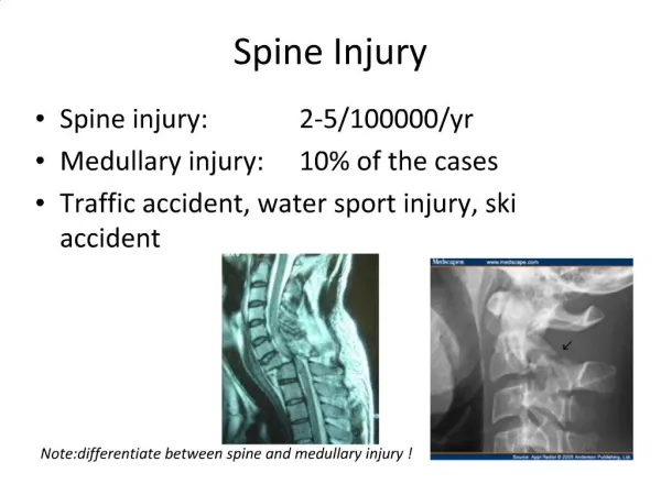
The curve of scoliosis often progresses most during the period of rapid skeletal growth and maturation. Serial x-rays show how this curve increased over a period of 4 years.
Reasons to treat • Cosmetic mainly. • Progression (>50 degree) as a rate of 0.5-1 /year. • Cardiac and respiratory complications. • Disc herniation. • Degenerative changes. • Patients with severe chest deformities should undergo pulmonary function tests. • A marked reduction in vital capacity is associated with diminished life expectancy and carries obvious risks for surgery.
Treatment • (Exercise – has no effect on the curve) • Patients with a curve <20 degree will not progress any more • Patients with a curve >50 degree may progress more →surgery is needed. • Those between 20-50→ need follow up • If the curve increases with time,treat as “B”,if doesn't ,considered as <20
Treatment "The 3O's ": (1) observation: 4-6 monthly visits. (2) Orthotics: braces, prevents progression and does not correct deformity. (3) Operative intervention.
Kyphosis • is an abnormally excessive convex kyphotic curvature (flexible or fixed) of the spine as it occurs in the cervical, thoracic and sacral regions. • A normal thoracic spine have a slight kyphotic angle(20° to 45°) When the"roundness" of the upper spine increases past 45° it is called kyphosis or "hyperkyphosis". • Common in males
Causes of kyphosis • degenerative diseases such as arthritis • developmental problems - Scheuermann's disease • osteoporosis with compression fractures of the vertebra • multiple myeloma or trauma.
Kyphosis types • Postural kyphosis • is common (‘round back’ or ‘drooping shoulders’) • may be associated with other postural defects such as flat-feet. • If treatment is needed, this consists of postural exercises. • Structural kyphosis • Osteoporosis of the spine (the common round back of elderly people) • Ankylosingspondylitis • Scheuermann’s disease (adolescent kyphosis).
A kyphos (or gibbus) • is a sharp posterior angulation due to localized collapse or wedging of one or more vertebrae. • Causes • a congenital anomaly • a fracture (sometimes pathological) • spinal TB.
Congenital kyphosis Type I : failure of formation • is the commonest (and the worst) type. • may lead to cord compression. Type II : failure of segmentation • The risk of neurological compression is much less.
Adolescent kyphosis(Scheuermann’s disease) This is a ‘developmental’ disorder Abnormal ossification (and possibly some fragmentation) of the ring epiphyses on the upper and lower surfaces of vertebral body in the growing spine. As a consequence these cartilaginous end-plates are weaker than normal and the affected vertebrae in the thoracic spine, may become wedge shaped. If this happens, the normal kyphosis is exaggerated. Schmorl’s nodes : small central herniations of disc material into the vertebral body.
Thoracic Scheuermann’s disease • usually, appears in the midthoracic vertebrae. • starts at or shortly after puberty • more common in males • The patient may complain of backache and fatigue.
Thoracic Scheuermann’s disease Diagnosis • X-rays : AP and lateral spine. Findings: • Anterior wedging • Disc narrowing • Endplate irregularities • Schmorl’s nodes • Scoliosis • Compensatory hyperlordosis • MRI: To rule out associated abnormalities of spinal curve and nerves. .
Thoracic Scheuermann’s disease Treatment • physiotherapy • If there is concern about back pain and/or deformity, an extension brace worn for 1 year • If this fails operative correction and fusion may be needed