
心电触发及门控
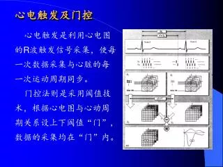
心电触发及门控. 心电触发是利用心电图的 R 波触发信号采集,使每一次数据采集与心脏的每一次运动周期同步。 门控法则是采用阈值技术,根据心电图与心动周期关系设上下阈值“门”,数据的采集均在“门”内。. 心电触发及门控. 参数设定 心动周期 (Hp)=60x1000/ 心动频率 (Hf) 触发延迟时间 Td, 为 R 波至开始采集的间隔时间。 T1 加权像,有效 TR 为一个 Hp ,一般 TR 为 Hp 小10%左右,防止心律不齐。 T2 加权时, TR 应为 二至三个心动周期。. 用颈的 CTL 阵列定相线圈 : 2D TOF 颈动脉 (GE).

心电触发及门控
E N D
Presentation Transcript
心电触发及门控 心电触发是利用心电图的R波触发信号采集,使每一次数据采集与心脏的每一次运动周期同步。 门控法则是采用阈值技术,根据心电图与心动周期关系设上下阈值“门”,数据的采集均在“门”内。
心电触发及门控 参数设定 心动周期(Hp)=60x1000/心动频率(Hf) 触发延迟时间Td,为R波至开始采集的间隔时间。 T1加权像,有效TR为一个Hp,一般TR为Hp小10%左右,防止心律不齐。 T2加权时,TR应为 二至三个心动周期。
肾脏MRA • 以钆增强三维MR血管造影术,用于代替导管血管造影术。血管呈高信号,MRA是一安全,快速和舒适的诊断方法,成本是通常血管造影术的三分之一。 • 肾脏动脉狭窄或动脉闭闭塞评价 • 为肾脏的移植评价 • 肾脏fibromuscular发育异常评价 右侧肾动脉在二裂处严重狭窄。扫描时间为30分钟左右
CE-MRA,MIP 无右侧椎动脉
CE-MRA,MIP fibromuscular dysplasia发育异常around right renal artery
三维TOF MRA:从动脉拱到威利斯环左侧颈动脉狭窄
Circle of Willis 左、右侧内部颈动脉(ICA)和基部动脉组成威利斯环向大脑向脑提供血液。 由于ICA受阻,通过环重新分配血液向颅脑提供血液。它的功能取决于环结构的连贯性。
Circle of Willis morphology anterior cerebral arteries(ACA) internal carotid artery (ICA) anterior communicating artery (ACoA) posterior cerebral arteries (PCA) Posterior communicating arteries (PCoAs) precommunicating segments of ACA (A1) Precommunicating segments (P1) of PCA
Circle of Willis morphology 1. 单、双侧显著狭窄70–99% 2. 单侧闭塞 3.单侧闭塞,并邻侧狭窄 4. 双侧闭塞.
Anatomical variations in the anterior part of the circle of Willis:
Anatomical variations in the posterior part of the circle of Willis
3D-TOF MR angiogram in a 24 year old man shows an entirely complete circle with anterior variant b and posterior variant a
MR angiogram in a 21 year old man shows a partially complete circle. Only the anterior part is complete, classified as an anterior variant a and a posterior variant e. The anterior choroidal arteries should not be mistaken for PCoAs.
MR angiogram in a 23 year old woman shows a partially complete circle. Only the posterior part is complete, with anterior variant g and a posterior variant a.
MR angiogram in a 63 year old woman with an entirely incomplete circle with anterior variant g and posterior variant d. The left PCoA is only visible on the source images: its signal intensity is too low for it to show up on the MIP.
Long-Axis Localizer • 估计LV和RV心肌的功能 • 主冠形状和三尖的回流 • 中隔损害,通常通过血液流动像来判断.
Short-Axis Localizer LV壁运动估价 LV和RV容量计算 喷射量 Base to Apex
Radial Viewer 左室像,心房概貌。 左室流出轨迹,主动脉回流。
Coronal Ascending Aorta Plane 升主动脉面评价
Single plane of 3D image set visualizes left main, LAD and circumflex coronary arteries. Acquisition done with non breath-hold, spiral technique.
2D Vessel Tracking using SmartPrep technique demonstrating RCA
Thirty second, breath-held pulmonary MRA demonstrating anomalous left pulmonary venous