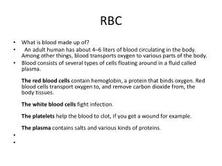
RBC
RBC. Qinshi Pan. Clinical Measurements: Hematocrit : RBC mass as % of blood volume (high as baby, dips at few months then men: 38.8-50/ female: 34.9-44.5) Hemoglobin: Total Hb / volume blood Hct/3= Hgb Red Cell Distribution (RDV): measure of anisocytosis

RBC
E N D
Presentation Transcript
RBC Qinshi Pan
Clinical Measurements: Hematocrit: RBC mass as % of blood volume (high as baby, dips at few months then men: 38.8-50/ female: 34.9-44.5) Hemoglobin: Total Hb/ volume blood Hct/3=Hgb Red Cell Distribution (RDV): measure of anisocytosis Low Hb and Hct (amt of RBC)= anemia Low MCV (size)= microcytosis Low MCH (color)= hypochromia Hyperchromic does not exist Haptoglobin: protein that binds plasma circulating heme, in hemolysis taken out of circulation (acute phase reactant) therefore hemolysis= less haptoglobin Hematopoiesis: SC RBC/WBC/platlets-can increase 4-5 fold in 7-10 days-usually in BM, can also be in liver, spleen, LNRBCs in intravascular space (not stored)O2 in kidneyEPO(peritubular caps) SC (clusters of dark cells) RBC 40% of blood RBC: transport O2 and CO2 for 100-120 days Aging: smaller, less elastic, spherical (removed in spleen) 4 factos: Normal vs impaired Hb Acute vs Chronic loss Extent of RBC volume loss Indirect effects (Fe, spleen, etc) • Hemolysis: • Intravascular (sudden, catastrophic): destruction of RBCs in BVsschistocytes, anemia, Hb goes up, Haptoglobin goes down, bilirubin increases, hemoglobinemia, hemoglobinuria Renal failure, DIC • - Immunehemolytic RBC, C’ mediated, severe osmotic stress • Extravascular(slow): chronic, enhancement, amp, of normal physiologic removal of RBCs anemia, elevated EPO, BM hyperplasia (rxn to decreased RBCs) Anemia:Hb or Hct<2.5 percentile after being adjusted for age, sex, machine, altitude [therefore clinical definition]4 approaches: Etiology, RBC size/morphology, frequency of occurrence, practical (Fe/B12 shot)
Normal Rouleux Echinocytes: may be artifact from storing Heinz body: Denatured hemoglobin Anisocytosis (size) Poikilocytosis (Shape) Basophilic Stippling: RNA Stomacytosis Pappenheimer body: Iron Howell-Jolly: DNA Spherocytosis Teardrop Elliptocytosis
Intrinsic Defects Congested Spleen Peripheral Blood Etiology: Blood Loss - Acute: trauma (normal hgb/hct) 10-15% loss in <1hr: S&S from defect in vascular volume not lack of O2 carrying capacity 20% loss in <1 hr: shock, postural hypoTN - Chronic: GI/Gyn: gradual b/c BM can increase production by 4-5 fold. S&S if Hb<7 Increased Destruction of RBCs - Intrinsic (Defective RBC): -Hereditary:spherocytosis, G6PD thalassemia, sickle cell anemia - Acquired: paroxysmal nocturnal hemoglobinuria - Extrinsic (Normal RBC): AIHA, mechanical trauma, Pb poisoning Impaired Red Cell production Hereditary Spherocytosis (memH): usu AD Ankryn, can also be spectrin. Unstable membrane shear stress in circulation loss of fragments S&S: 6-9 Hb, spleno, cholelithiasis Dx: osmotic fragility test Rx: Splenonectomy Sequelae: parvo infection aplastic crisis Paroxysmal Nocturnal Hemoglobinuria (memA): GPI anchor mutless CD55/59/C8BP C’ med chronic hemolysis WITHOUT severe hemoglobinuria acute at night S&S:dark urine (morning), hemolysis and hemoglobinuria Ass: venous thrombosis, AML
Thalassemia (Quantitative): 2 beta and 2 alpha globin make 1 Hb PB: anisocytosis, microcytosis, Crewcut radiograph Alpha: SE Asia Chrom 16 (a1-4): -/a a/a: silent carrier, asymptomatic -/- a/a or -/a -/a: asympt like Bminor -/- -/a: HbH disease, severe like Binter -/- -/-: HydrobsFetalis: die in utero Beta:mediterranian Chrom 11 (B1-2): B- (normal) B0/B+ (mutation) B0/B0 or B+/B+ or B0/B+: Major, Severe, transfuse Variable: Intermediate, severe but don’t need to transfuse B0/B-, B+/B-: Minor, asymptomatic G6PD Deficiency (metabH): XR can’t protect as well from free radicals GDPD A-: blacks, moderately reduced GDPD Mediterranean: Middle east, Fava bean hemolytic episode Both protect against Malaria. Patho: oxidative stress acute intravascular hemolysis anemia, hemoglobinuria, hemoglobinemia removed extravascularily PB: Heinz bodies and bite cells Episodes are self-limited if stress is removed Intrinsic Defects Sickle Cell Disorders (Qualitative): 6 position of beta GluVal(Hetero: trait, Homo:Disease) 5-6mo (no HbF) Sickle crisis (severe bone/lung/liver/brain/penis pain)/ Autosplenectomy (esp with S pneumo and H. Flu), also see gallstones, skin ulcers 50% to 50yo Dx: Hb electrophoresis, DNA testing, HbS solubility Treat with hydroxyurea Spleen cong. w/ sickle cells Sickle cell
Extrinsic Defects Anemia caused by impaired RBC production Decreased(Diminished) or Ineffective Erythropoiesis • Megaloblastic: B12 and folatedeficiency (nutritional) • Iron Deficiency (nutritional) • Anemia of Chronic Disease (inhibits hematopoeisis) • Aplastic anemia and pure red cell aplasia (destroy SCs) Marrow failure: replacement/displacement marrow space • Myelofibrosis, primary versus secondary • Space-occupying • Hematologic malignancy • Metastatic non-hematologic malignancy Chemical Toxicity (Pb): Patho: Pb binds sulfhydryl groups in ferrochelatase displaces iron amd makes zinc protoporphyrin/erytrocyteprotoporphyrin PB: hypochro, microcytic anemia, basophilic stippling Traumatic Hemolytic Anemia: Path: trauma RBCs turn to fragments intravascular hemolysis Mechanical: prostethic cardiac valve Microangiopathic: TTP/HUS/DIC Immunohemolytic: Warm (IgG): idiopathic, SLE, drugs, CLL Severe, life threatening extravascularhemolysis (hard to treat b/c no compatible blood) Cold (IgM): mycoplasma, mono (younger, abrupt, severe) idiopathic, Waldenstrom, CLL, DLBCL, splenic lymphoma (older, mild, worse when cold) Cold (IgG): Cold hemolysin Hemolytic Anemia paroxysmal cold hemoclobinuria. RARE
Megaloblastic Anemia (B12 and Folate): B12 Deficiency: elevated homocysteine and methylmalonic acid (better sensitivity than decreased serum cobalamin) Pernicious Anemia: Patients have antiparietal cell Ig No IF no Bwe S&S: glossits, gastric atrophy, neurological (can occur before B12 goes too low) cortex/lateral columns Usually 60 Other causes: Nutritional, malsorption, competitive uptake by parasites, increased requirement (pregnancy/ hyperthyroidism) PB: hypersegmentednp with 6 lobes + leukopenia, severe macrocyticanemia, hyperbilirubinemia (extravascualrhemolysis) BM: ineffective erythropoiesis Dx: serum B12, MMA, homocysteine (also up in B9 deficiency), Schillings test, pernicious needs Ig test Folic Acid: common in alcoholics, pregnant, and those taking methotrexate. Does not present with neurological symptoms • Iron Deficiency: • insufficient intake (diet, malsorption, celiac, removal of ileium (Chrons), secondary to systemic • excessive loss (blood loss, hemodialysis) • excessive use (growing children, pregnancy, lactation) • S&S: smooth tongue, spoon nail • PB:hypochromatic, microcytic RBC, low serum ferritin/iron/transferrin saturation/stores, increased iron-binding capacity • Dx: Iron studies then look at reticulocytes to see if there’s BM problem • BMBx:Prussian Blue stain for Iron • Anemia of Chronic Disease: Decreased EPO or can’t move Fe to erythroid precursor, MCC in hospitalized patients besides post-surgical and acute hemorrhage • Chronic infections: osteomyelitis/endocarditis • Immune (RA, Chrons) • Malig: Hodkin, Carcinoma of lung/breast • High serum ferritin (Fe def has low) • Definitive: BM has prussian blue macrophage • Aplastic Anemia: idiopathic, chemo (alkylatin), Chloramphenicol (ABX), Idiosyncratic, physical agents (Hep, CMV, VZ, fanconi, telomerase) • FATTY BM- think back to WBC • Myelophthsic Anemia: like aplastic but instead of messing with RBCs you have a mass in the bone
Blood Testing • ABO and RhD • Hep B/C, HIV, HTLV, Syphilis • Acute hemolytic Anemia: • Burning along vein, low back pain, chills, fever, shock, increase pulse rate, DIC • Lab: increased bilirubin and LDH • Febrile, Nonhemolytic Reaction • Increase in temp by 1 degree, chills • Allergic Reaction: • Urticaria, pruritis, facial/glottal edema • Anaphylactic Reaction: • ANS dysregulation, dyspenea, pulmonary edema, bronchospasm, hypotension • Transfusion Related Acute Lung Injury (TRALI) • Acute respiratory distress in 6 hrs with hypoxemia and bilateral pulmonary infiltrates • ABO Compatibility: • O can only get O (universal donor) • AB can get every type (universal acceptor/ blood hogger) • A can’t have B, B can’t have A Polycythemia: RBC mass > 97.5 percentile (remember to adjust for altitude, age, sex) Erythrocytosis: Increased RBCs with no increase in WBC or platelets MCC: smoking, high alt., blue bloaters Polycythemiarubravera: see WBC notes (comes with increase in WBC and platlets) -- increased viscosity, CV complications, thrombosis, hemmorrhage Dx: phlebotomy • Blood Products • Platelets: replaces platlets • 10,000/ml if stable, >20,000/ml if unstable or > 50,000/ml in bleeding or surgical patients • Frozen Plasma: replace coagulation factors • active bleeding or massive transfusions, emergency reversal warfarin effect, • Cryoprecipitatedantihemophilic factor • Replace fibrinogen and Factor XIII • Replace factor VIII or vWF (if factor VIII and factor VIII/vWF • Cellular Therapy Products • White cells • Stem cells from bone marrow, cord blood, or apheresis