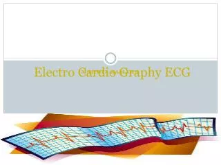
Electro Cardio Graphy ECG
Electro Cardio Graphy ECG. Mahdia Samaha . Definition of ECG. The ECG is a graphic representation of the electrical impulses that the heart generates during the cardiac cycle.

Electro Cardio Graphy ECG
E N D
Presentation Transcript
Electro Cardio Graphy ECG Mahdia Samaha
Definition of ECG • The ECG is a graphic representation of the electrical impulses that the heart generates during the cardiac cycle. • These electrical impulses are conducted to the body's surface, where they are detected by electrodes placed on the patient's limbs and chest. • The monitoring electrodes detect the electrical activity of the heart from a variety of spatial perspectives. • The ECG lead system is composed of several electrodes that are placed on each of the four extremities and at varying sites on the chest. Each combination of electrodes is called a lead.
12-lead ECG • It provides a comprehensive view of the flow of the heart's electrical currents in two different planes. • There are six limb leads (combination of electrodes on the extremities) and six chest leads (corresponding to six sites on the chest). • standard limb leads • Leads I: records the difference in electrical potential between the left arm (LA) and the right arm (RA). • Lead II: records the electrical potential between the RA and the left leg (LL). • Lead III reflects the difference between the LA and the LL. The right leg (RL) electrode is an inactive ground in all leads.
Augmented limb leads • aVR • aVL • aVF • The augmented leads measure the electrode potential between the center of the heart and the right arm (aVR), the left arm (aVL), and the left leg (aVF).
Limb leads • Rt arm (avr) Red color. • Lt arm (avl) Yellow color. • Lt leg (avf) Green color. • Rt leg, black color.
Chest, or precordial leads • The six standard, (V1, V2, V3, V4, V5, V6) are placed at six different positions on the chest, surrounding the heart. • In general, it is said that leads II, III and aVF look at the inferior part of the heart, leads aVL and I look at the lateral part of the heart, and leads V2-V4 look at the anterior part of the heart.
Steps to reading ECGs • What is the rate? Both atrial and ventricular if they are not the same. • Is the rhythm regular or irregular? • Do the P waves all look the same? Is there a P wave for every QRS and conversely a QRS for every P wave? • Are all the complexes within normal time limits? • Name the rhythm and any abnormalities.
ECG recording Small and Large Boxes on EKG Trace On monitoring paper, each small square represents .04 seconds. .04 seconds One large square five small boxes on each axis. .04 x 5 = .20 seconds
ECG waves • P wave: This represents atrial electrical depolarization associated with atrial contraction. It represents electrical activity associated with the spread of the original impulse from the sinoatrial (SA) node through the atria. • PR interval: This represents the time required for the impulse to travel from the SA node to the atrioventricular (AV) node. If prolonged PR interval: a conduction delay exists in the AV node (e.g., a first-degree heart block). If the PR interval is shortened: the impulse must have reached the ventricle through a "shortcut" (as in Wolff-Parkinson-White syndrome).
ECG waves • QRS complex. This represents ventricular electrical depolarization associated with ventricular contraction. This consists of: • initial downward (negative) deflection (Q wave) • a large upward (positive) deflection (R wave) • a small downward deflection (S wave). A widened QRS complex: indicates abnormal or prolonged ventricular depolarization time (as in a bundle-branch block).
ECG waves • ST segment. This represents the period between the completion of depolarization and the beginning of repolarization of the ventricular muscle. This segment may be elevated or depressed in transient muscle ischemia (e.g., angina) or in muscle injury (as in the early stages of myocardial infarction). • T wave: This represents ventricular repolarization (i.e., return to neutral electrical activity). • U wave: This deflection follows the T wave and is usually quite small. It represents repolarization of the Purkinje nerve fibers within the ventricles
Interfering factors • Inaccurate placement of the electrodes • Electrolyte imbalances • Poor contact between the skin and the electrodes • Movement or muscle twitching during the test • Drugs that can affect results include digitalis, quinidine, and barbiturates
Abnormal findings • Arrhythmia • Acute myocardial infarction • Myocardial ischemia • Old myocardial infarction • Conduction defects • Conduction system disease • Wolff-Parkinson-White syndrome • Ventricular hypertrophy • Cor pulmonale • Pulmonary embolus • Electrolyte imbalance • Pericarditis
Rate • Look at complexes in a 6-second strip and count the complexes; that will give you a rough estimate of rate • Count the number of large boxes between two complexes and divide into 300 • Count the number of small boxes between two complexes and divide into 1500 • Estimate rate by sequence of numbers (see next slide)
Normal Timing • PR interval – 0.12 to 0.20 seconds • QRS interval – less then 0.12 • QT interval – varies with rate. It is usually less then ½ the R-to-R distance on the preceding waves
Normal Sinus Rhythm • Rate = 60-100 beat / minute. • The rhythm is regular • All intervals are within normal limits • There is a P for every QRS and a QRS for every P. • P : QRS ratio = 1 : 1. • The P waves all look the same • Presence of P, QRS, T in each cycle. • Normal shape, time of waves, segments and intervals
Arrhythmias • Definition of arrhythmias: any disturbance in the rate, regularity, site or origin, or conduction of cardiac electrical impulse. It can be single beat or sustained rhythm. • Clinical Manifestaions: • Palpitation. • Light headedness and syncope R/T low cardiac output. • Angina R/T increased O2 demand. • Sudden death.
Arrhythmias Causes: [ HISDEBS ] • H = Hypoxia. • I = Ischemia. • S = Sympathetic stimulation. • D = Drugs. • E = Electrolyte disturbances. • B = Bradycardia. • S = Stretch = cardiac enlargement
Sinus Arrhythmia • Rate is between 60 and 100 beats/minute • The rhythm is irregular. The SA node rate can increase or decrease with respirations • All intervals are within normal limits • There is a P for every QRS and a QRS for every P • The P waves all look the same • More common in children and athletes • Ask the patient to stop breathing and the rate will become regular
Sinus Tachycardia • Rate above 100 beats/minute • The rhythm is regular • All intervals are within normal limits • There is a P for every QRS and a QRS for every P • The P waves all look the same Caused by: fever, stress, caffeine, nicotine, exercise, or by increased sympathetic tone Treatment: to take care of the underlying cause
Sinus Bradycardia • Rate is lower than 60 beats/minute • The rhythm is regular • All intervals are within normal limits • There is a P for every QRS and a QRS for every P • The P waves all look the same Caused by; beta-blocker, digitalis, or calcium channel blockers. Normal for athletes • Don’t treat unless there are symptoms. Can use pacing or atropine
Super-ventricular Arrhythmia • Premature Atrial Contraction PAC. • Junctional Pre-mature beats. • Atrial Flutter. • Atrial fibrillation
Premature Atrial Contraction (PAC) • Originate in the atria. • Neither indicate disease nor requiring treatment. • P wave differ in shape and time from normal P (introduces it-self before the next anticipated sinus wave. • Can occur at any rate • The rhythm is irregular because of the early beat but is regular at other times • All intervals can be within normal limits • There is a P for every QRS and a QRS for every P • The P waves all look the same except the P in front of the PAC will be different
Atrial Flutter • Ventricular and atrial rate: Atrial rate ranges between 250 and 400; ventricular rate usually ranges between 75 and 150. • The rhythm is regular or regularly irregular • There is no PR interval. QRS may be normal • P: QRS ratio: 2:1, 3:1, or 4:1 • There are no P waves; they are now called flutter waves = saw tooth pattern • Problem: Loss of atrial kick and ventricular conduction is too fast or too slow to allow good filling of the ventricles
Atrial Fibrillation • Atrial rate is between 350 and 600 beats/minute; ventricular rate can vary 120-180 b/m • The rhythm is irregular • No P waves. • There is no PR interval; QRS may be normal • There are many more f waves then QRSs • Unlike flutter where the f wave will appear the same, in fib the f waves are from different foci so they are different • Caused by mitral valve disease or CAD. • Carotid massage slow the ventricular contraction.
Function at the Junction • Junctional or idionodal rhythm occurs when the AV node, instead of the sinus node, becomes the pacemaker of the heart. • When the sinus node slows (eg, from increased vagal tone) or when the impulse cannot be conducted through the AV node (eg, because of complete heart block), the AV node automatically discharges an impulse.
Junctional Rhythm • Ventricular and atrial rate: Ventricular rate 40 to 60; atrial rate also 40 to 60 if P waves are discernible • Ventricular and atrial rhythm: Regular • QRS shape and duration: Usually normal, but may be abnormal • P wave: May be absent, after the QRS complex, or before the QRS; may be inverted, especially in lead II • PR interval: If P wave is in front of the QRS, PR interval is less than 0.12 second. • P: QRS ratio: 1:1 or 0:1
Atrioventricular Nodal Reentry Tachycardia • Occurs when an impulse is conducted to an area in the AV node that causes the impulse to be rerouted back into the same area over and over again at a very fast rate. • Each time the impulse is conducted through this area, it is also conducted down into the ventricles, causing a fast ventricular rate.
Paroxysmal atrial tachycardia • AV nodal reentry tachycardia that has an abrupt onset and an abrupt cessation with a QRS of normal duration had been called paroxysmal atrial tachycardia (PAT). • Factors associated with the development of AV nodal reentry tachycardia include caffeine, nicotine, hypoxemia, and stress.
Atrioventricular Nodal Reentry Tachycardia • Ventricular and atrial rate: Atrial rate usually ranges between 150 to 250; ventricular rate usually ranges between 75 to 250 • Ventricular and atrial rhythm: Regular; sudden onset and termination of the tachycardia • QRS shape and duration: Usually normal, but may be abnormal • P wave: Usually very difficult to discern • PR interval: If P wave is in front of the QRS, PR interval is less than 0.12 seconds • P: QRS ratio: 1:1, 2:1
If P waves cannot be identified, the rhythm may be called supraventricular tachycardia (SVT), which indicates only that it is not ventricular tachycardia (VT). • SVT could be atrial fibrillation, atrial flutter, or AV nodal reentry tachycardia, among others. • Vagal maneuvers and adenosine are used to slow conduction in the AV node to allow visualization of the P waves.
Junctional Arrhythmia • Rate is between 40 and 60 beats/minute • The rhythm is regular • There is a P for every QRS and a QRS for every P • The P wave can be in three possible places • Retrograde conduction to atria before ventricle; P wave would be upside down before the QRS • If both atria and ventricle receive stimulus at the same time, the P would be buried in the QRS • If the ventricle was stimulated first, the P would be located just after the QRS
Junctional Rhythms • Junctional bradycardia • Rate less than 40 beats/minute • Accelerated junctional • Rate 60-100 beats/minute • Junctional tachycardia • Rate is greater then 100 beats/minute • Premature junctional contractions (PJC) • Early beats in the cycle that have junctional P wave morphology
Ventricular Arrhythmia Premature ventricular contractions PVCs. Ventricular tachycardia. Ventricular fibrillation.
Premature Ventricular Contractions (PVC) • Early beat that is wide (>0.12) • Originates the ventricles • No P wave • Compensatory pause • Can be defined by couplet or triplet; anything more would be considered ventricular tachycardia • Monomorphic or polymorphic, R on T Treat if > 6 PVCs/min, couplets or triplets, multifocal, or R on T. If not treated cause VT and VF. Lindocaine is the Rx. Of choice.
Premature Ventricular Contractions (PVC) • Premature ventricular complex • (PVC) is an impulse that starts in a ventricle and is conducted through the ventricles before the next normal sinus impulse. • PVCs can occur in healthy people, especially with the use of caffeine, nicotine, or alcohol. They are also caused by cardiac ischemia or infarction, increased workload on the heart (eg, exercise, fever, hypervolemia, heart failure, tachycardia), digitalis toxicity, hypoxia, acidosis, or electrolyte imbalances, especially hypokalemia.
Premature Ventricular Contractions (PVC) • Ventricular and atrial rate: Depends on the underlying rhythm (eg, sinus rhythm) • Ventricular and atrial rhythm: Irregular due to early QRS, creating one RR interval that is shorter than the others. PP interval may be regular, indicating that the PVC did not depolarize the sinus node. • QRS shape and duration: Duration is 0.12 seconds or longer; shape is bizarre and abnormal • P wave: Visibility of P wave depends on the timing of the • PVC; may be absent (hidden in the QRS or T wave) or in front of the QRS. If the P wave follows the QRS, the shape of the P wave may be different. • PR interval: If the P wave is in front of the QRS, the PR interval is less than 0.12 seconds. • P: QRS ratio: 0:1; 1:1
Ventricular Tachycardia • Rate is between 100 and 200 beats/minute • The rhythm is regular, but can change to different rhythms • No PR interval; QRS is wide and aberrant • There may be a P wave, but it is not related to the QRS