Download
1 / 32
320 likes | 414 Views
RESPIRATORY SYSTEM. Functions of the Respiratory System. Take in oxygen Get rid of carbon dioxide Helps with smelling Filters air that is inhaled Produces sounds Rids the body of some water and heat in exhaled air. Organs of the Respiratory System.

E N D
Functions of the Respiratory System • Take in oxygen • Get rid of carbon dioxide • Helps with smelling • Filters air that is inhaled • Produces sounds • Rids the body of some water and heat in exhaled air
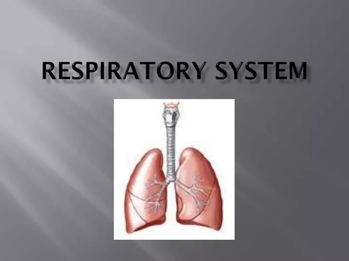
Organs of the Respiratory System The organs of the respiratory system include the nose, pharynx, larynx, trachea, bronchi, bronchioles, alveoli and lungs. Nose: • The 2 external openings are called external nares. • The internal portion of the nose has 2 openings that connect to the throat called internal nares • The nose is divided into right and left internal parts by the septum.
Functions of the Nose • Coarse hairs in the nose filter out large dust particles • Nasal conchae- 3 shelves that extend out of the wall of the nasal cavity to increase surface area for warming, moistening and filtering. • Mucous membranes line the nasal cavity and secrete mucus that moistens the air and traps dust particles. • Above the conchae is the olfactory epithelium which detects smells.
Pharynx and Larynx • Pharynx is also called the throat- A tube that starts at the internal nares and extends part way down the neck. • A passageway for air and food and provides a resonating chamber for speech sounds. • Larynx is the voice box. It is a short passageway that connects the pharynx with the trachea. • Thyroid cartilage (Adam’s apple) forms the anterior wall of the larynx • Epiglottis is elastic cartilage that is the most superior part of the larynx. It prevents food and drink from entering the larynx.
Voice Production • The mucous membrane of the larynx forms 2 pairs of folds: false vocal cordsand true vocal cords. • True vocal cords produce sound. They are elastic ligaments that are stretched between pieces of cartilage. They are also attached to muscles that pull the ligaments tightly when contracted. This moves the true vocal cords out into the air passageway. Air pushed against the cords causes them to vibrate and produce sound waves. • Males have thicker vocal cords due to male sex hormones. These vibrate more slowly and produce a deeper pitch.
Trachea • Windpipe- Air passageway anterior to the esophagus • Extends from the larynx to the 5th thoracic vertebra • Wall is lined with mucous membrane • Cilia also line the wall-move mucus up the trachea • Supported by cartilage- 16 to 20 C-shaped rings of hyaline cartilage stacked on top of another.
Trachea obstruction • Heimlich maneuvermay be used to expel an aspirated object. • If not successful, a tracheostomymay be performed- incision in the trachea below the cartilage and a tube is inserted. • Intubationis another method when a tube is inserted into the mouth or nose and passed down through the larynx and trachea.
Bronchi • Bronchiare branches of the trachea that each feed a lung. • The right bronchus is more vertical, shorter and wider than the left. As a result, foreign objects are more likely to enter and lodge in the right bronchus than in the left. • Bronchi contain C-rings of cartilage and are lined with cilia. • Bronchi branch into smaller bronchioles, which do not contain cartilage rings but are lined with more smooth muscle for support.
Asthma • Asthma is chronic airway inflammation due to hypersensitivity to a variety of stimuli. These triggers include allergens, emotional upset, exercise, breathing cold air and cigarette smoke. • Triggers cause the walls of the bronchi and bronchioles to spasm, the mucous membranes to swell, increased mucus secretion or damage to the lining of an airway. • Asthma symptoms include difficulty breathing, coughing, wheezing, chest tightness, fatigue and anxiety.
Lungs • Lungs are covered directly with visceral pleura and the lung cavity is lined with parietal pleura. • Pleurisy- inflammation of the pleural membranes which causes friction during breathing which is quite painful. • The narrow top of each lung is the apex. The broad bottom is the base. The area on the medial side through which bronchi enter is called the hilus. • The right lung has 3 lobes and the left lung has 2 lobes. The left lung has a concavity where the heart lies- called the cardiac notch.
Alveoli • The airways dead-end with thin air sacs called alveoli. • Associated with alveoli walls are alveolar macrophages which remove fine dust particles and other debris. • Capillaries surround the alveoli and allow for gases to be exchanged between the respiratory tissue and the blood.
Lung Volume • Normal quiet breathing is called eupnea. • A cessation in breathing is called apnea. • Labored breathing is called dyspnea. • At rest, a healthy adult takes 12 breaths a minute. • The volume of one breath is called tidal volume. • Minute ventilation is the total volume of air inhaled and exhaled each minute. • MV=12 breaths/min X 500 mL/breath = 6 liters/min
Ventilation • The 4 basic events of respiration are: ventilation, external respiration, internal respiration, and transport of respiratory gases. • Ventilation- The passive process by which air flows into and out of the lungs. This occurs due to differences in air pressure between the thoracic cavity and the atmosphere. - Inhalation- Air rushing in- occurs when the atmospheric air pressure is greater than the thoracic cavity air pressure. • What causes the thoracic air pressure to decrease?
Pressure Changes • Thoracic air pressure decreases and inhalation occurs due to: • Contraction of the diaphragm (flattening) • Contraction of muscles that cause the ribs and sternum to elevate anteriorly. • Expansion of the lungs due to the parietal and visceral pleura being pulled outward. • Exhalation- Air rushing out- occurs when the thoracic cavity air pressure is greater than the atmospheric air pressure. This pressure difference is due to: • Elastic recoil of the chest wall and lungs • Relaxation of the diaphragm
External Respiration • External Respiration is the exchange of O2 and CO2 between the air in the alveoli and the blood in the capillaries around the alveoli. • Internal Respiration is the exchange of gases between the blood in the capillaries and the cells of the body. • What causes these gases to be exchanged? • Diffusion- movement of molecules from where there is more of them to where there is less of them. • O2 moves from the alveoli (higher concentration) to the capillaries (lower concentration) • CO2 moves from the capillaries (higher concentration) to the alveoli (lower concentration)
Transport of Respiratory Gases • Oxygen does not dissolve well in water. Therefore, only 1.5% of oxygen dissolves in plasma. Most of it is carried by hemoglobin of erythrocytes. • About 7% of carbon dioxide dissolves in plasma. About 23% binds with hemoglobin. About 70% of carbon dioxide is transported in the bloodstream(plasma) as bicarbonate ions- HCO3-
Carbon Monoxide Poisoning • CO is an odorless and colorless gas produced when there is incomplete burning of natural gas or any carbon containing fuel. • Why is it dangerous when inhaled?
CONTROL OF RESPIRATION • At rest, 200 ml of oxygen are used by the body’s cells each minute. Of course, this need is increased during exercise. • There are groups of neurons in the brain stem which adjust respiratory effort to meet metabolic demand. These neurons are called the respiratory center. • The 3 parts to the brain stem include the midbrain, pons and medulla oblongata.
Respiratory Center • There are 3 areas to the respiratory center: • Medullary rhythmicity area- Located in the medulla oblongata- This controls the basic rhythm of respiration. During quiet breathing, inhalation lasts 2 seconds and exhalation lasts 3 seconds. • Pneumotaxic area- Located in the upper pons- This turns off inspiration before the lungs become too full and pressurized. 3.Apneustic area- Located in the lower pons- This sends stimulatory impulses to increase and activate inhalation. This can be overidden by the pneumotaxic area.
Other Respiration Influences • Temperature- Increased temp = increased respiration. A sudden cold stimulus causes apnea- temporary cessation of breathing. • Pain- Sudden pain causes brief apnea and prolonged pain increases respiratory rate. • Stretching the anal sphincter muscle- Increases respiratory rate. This is sometimes done to newborns. • Blood pressure- A rise in blood pressure decreases respiration rate and vice versa.
Aging and Respiration • As we age, the following changes occur to the respiratory system: • Tissues become less elastic which results in a decrease of lung capacity. • There is a decrease in oxygen levels in the blood. • There is a decrease in the activity of alveolar macrophages • There is diminished ciliary action of the respiratory tract Because of these, elderly people are more susceptible to pneumonia, bronchitis, and emphysema.
BINGO 1. Apneustic center 2. 3. 4. 5. 6. 7. Septum 8. 9. 10. 11. 12. 13. Thyroid cartilage 14. 15. Olfactory epithelium 16. 17. 18. 19. Larynx 20. 21. 22. 23. 24.