Download

1 / 11
110 likes | 265 Views
The Basal Ganglia. Dr. Nimir Dr. Safaa. Objectives Understand the anatomical and functional definition of the basal ganglia. Identify the different components of the basal ganglia.

E N D
The Basal Ganglia Dr. Nimir Dr. Safaa
Objectives • Understand the anatomical and functional definition of the basal ganglia. • Identify the different components of the basal ganglia. • Describe the connections of the different components of the basal ganglia and the indirect pathways from the basal ganglia to the lower motor neurons. • Describe signs and symptoms of lesions which affect different components of the basal ganglia.
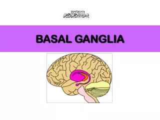
Basal ganglia are masses of gray matter situated deep within each cerebral hemisphere. • The basal nuclei play an important role in control of posture and voluntary movement, but have no direct input or output connections with spinal cord. • They are: • Corpus striatum. • Amygdaloid nucleus. • Claustrum. • SubstantiaNigra. • Subthalamic Nuclei.
Corpus striatum: • Is lateral to thalamus and divided by internal capsule, into caudate nucleus and lentiform nucleus. • Caudate nucleus has head, body & tail which terminates in amygdaloid nucleus. • Lentiform nucleus is lateral to internal capsule & medial to external capsule. • It is divided into dark putamen & light globuspallidus. • Neostriatum means putamen & caudate nucleus.
Amygdaloid nucleus: • Is in temporal lobe close to uncus. • It is part of the limbic system. • Claustrum: • Is thin gray matter lateral to external capsule and medial to subcortical white matter of insula. • Substantianigra and subthalamic nuclei: • Substantianigra of midbrain and subthalamic nuclei of diencephalon are functionally related to basal nuclei.
Connections: • Caudate nucleus and putamenare main sites for receiving input to the basal nuclei. • The globuspallidusis major site of output from basal nuclei. • Connections of the Corpus Striatum: • Afferent fibers: • Corticostriate. • Thalamostriate. • Nigrostriate. • Brainstem.
Efferent fibers: • Striatopallidal. • Striatonigral. • Connections of the globuspallidus: • Afferent: • Striatopallidal. • Efferent : • Pallidofugal Fibers which are: • Ansalenticularisto thalamic nuclei. • Fasciculus lenticularisto subthalamus. • Pallidosubthalamic to subthalamic nuclei. • Pallidotegmental to tegmentum of the midbrain.
Functions of the basal nuclei: • Basal nuclei control muscular movements by influencing cerebral cortex. • They have no direct control through descending pathways to brainstem and spinal cord. • They assist in regulation of voluntary movement and learning of motor skills.
Disorders of the basal nuclei: • Are of two types:Hyperkinetic disorders are those in which there are excessive and abnormal movements (chorea, athetosis, and ballism). • Hypokinetic disorders are those in which there is a lack or slowness of movement. • Parkinson disease includes both types of motor disturbances.