Download
1 / 1
10 likes | 244 Views
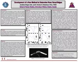
Figure 2. Instrumented lateral support. Biomechanics Laboratory. Development of a New Method to Determine Knee Varus/Valgus Jonathan Singer, B.Sc. and D. Gordon E. Robertson, Ph.D., FSCB School of Human Kinetics, University of Ottawa, Ontario, Canada. Introduction

E N D
Figure 2. Instrumented lateral support. Biomechanics Laboratory Development of a New Method to Determine Knee Varus/Valgus Jonathan Singer, B.Sc. and D. Gordon E. Robertson, Ph.D., FSCB School of Human Kinetics, University of Ottawa, Ontario, Canada Introduction Excessive knee varus/valgus is known to alter joint loading patterns, placing unusual stresses on the articular surfaces, joint capsule and ligaments. Such stresses can be the result of either a chronic condition, such as genu varum or valgum (Kerrigan et al., 2002), or an acute injury, which may occur during physical activity (Besier et al., 2001). Three-dimensional videography and inverse dynamics have been extensively used to quantify the varus/valgus moment at the knee. Such analyses can often be time consuming for the clinician, who must assess many patients, and difficult for the researcher, who may wish to set up equipment to collect data in remote locations. Methods Data from two subjects, performing a number of walking and crossover cutting trials, were collected with the participant wearing the instrumented brace. To ensure consistency among the cutting trials, the angle of cut was monitored, effectively falling between 30 and 60 degrees from the original direction of motion. Force platform data, using one Kistler force platform, were collected simultaneously with lateral- support strain gauge data for the stance phase of the left leg during one stride. All data were sampled at 240 Hz for the duration of stance and swing phases. Results and Discussion Figure 1 illustrates the moment of force pattern that was applied to the brace during one stride, beginning and ending at heel-strike. It is interesting to note that in the walking condition, the moment applied to the brace lacks the initial peak that has been reported to occur within the initial 15-30% of the stance phase (Allard et al., 1997). This may be due to the fact that the internal valgus moment of force effectively controls the varus motion of the leg during weight acceptance, resulting in a smaller applied moment to the brace. The second varus peak, occurring just prior to toe-off, corresponds with the internal valgus moment that typically occurs during 60-85% of stance (Allard et al., 1997). The moment applied to the brace in the crossover cut appears to be representative of that condition, whereby a greater moment is applied over the duration of stance. Purpose To develop a new method to determine the external knee varus/valgus moment. The proposed method employs strain gauges configured to measure the bending moment applied to the lateral support of a knee brace. Results and Discussion Peak varus moment of force values for the walking and crossover cut condition were 0.59 N·m and 0.67 N·m, respectively. Although the peak varus moment applied to the brace was larger in the crossover cut condition, it was not substantially so. This may be due to the relatively low coefficient of static friction (μ = 0.52) between the shoe sole and the force platform, barring the participant from sufficiently planting the foot and performing a vigorous cut. As the participants were aware of the need to perform a crossover cut, the small difference between the two applied moments may also be caused by postural adjustments made in preparation for the cut, in attempt to protect the knee structures (Besier et al., 2001). From this, future work entails comparing applied moments to internal knee moments and collecting data in vivo to account for concerns due to friction and anticipatory mechanisms. Methods Four strain gauges–two active and two Poisson–were wired in a Wheatstone bridge and applied to the distal aspect of the lateral support of a functional knee brace, such that the bending moment applied to the support could be measured. To determine the sensitivity of the lateral support, the most distal aspect was securely clamped in a vice and a range of known forces were applied to the support at a point just below the hinge. Applied moment of force values were correlated with the lateral support gauge output voltages. The sensitivity of the lateral support gauges, in both varus and valgus directions, was determined by the slope of the least squares regression line. Figure 1. External bending moment of force applied to the lateral support (top) and corresponding ground reaction forces (bottom) during walking (left) and crossover cutting (right). References Allard, P. et al. (1997). Three-dimensional Analysis of Human Locomotion. Toronto:Wiley. Besier, T.R. et al.(2001). Med Sci Sport Exer, 33:1176-81. Kerrigan, D.C. et al.(2002). Arch Phys Med Rehab, 83:889-93.


