
INTEGUMENTARY SYSTEM
SKIN Health Science Technology I Dr. Halbert. INTEGUMENTARY SYSTEM. Three main layers of skin. Epidermis. Dermis. Subcutaneous Fatty Tissue. Epidermis. Outermost layer Cells constantly being shed Contains no blood vessels or nerves 5 layers Stratum corneum: outer part

INTEGUMENTARY SYSTEM
E N D
Presentation Transcript
SKIN Health Science Technology I Dr. Halbert INTEGUMENTARY SYSTEM
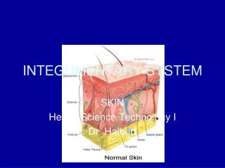
Three main layers of skin Epidermis Dermis Subcutaneous Fatty Tissue
Epidermis • Outermost layer • Cells constantly being shed • Contains no blood vessels or nerves • 5 layers • Stratum corneum: outer part • Stratum germinativum: inner layer
Dermis • “true skin” • Made up of elastic connective tissue and contains vessels, nerves, glands, hair follicles • Top layer covered with papillae which form ridges which make up our fingerprints
Subcutaneous fascia or hypodermis • Innermost layer • Connects skin to the muscle underneath
Sudoriferous glands • Sweat glands • Eliminate water, salts and some body wastes • Coiled tubes
Sebaceous glands • Oil glands • Produce sebum • Open in to hair follicles • When plugged results in pimple or blackhead
Alopecia • Lack of hair
Functions of the skin • Protection • Perception • Regulation of temperature • Storage of fat, water, vitamins • Absorption • Excretion • Production of Vitamin D
Skin pigment • Melanin: brown-black pigment, does absorb UV light resulting in a tan • Carotene: yellowish-red pigment
Albino • Absence of color pigment
Erythema • Reddish color of the skin • Burns or congestion of blood in vessels
Jaundice • Yellow discoloration • Liver or gallbladder disease or destruction of red blood cells
Cyanosis • Bluish discoloration • Insufficient oxygen
Macules • Flat spots on the skin • Ex: freckles
Papules • Firm raised areas • Pimples • Some stages of chicken pox
Vesicles • Blisters • Fluid filled sacs • Chicken pox
Pustules • Pus filled sacs • Ex: acne
Crusts • Dried pus and blood • scabs
Wheals • Itchy elevated areas with irregular shape • Hives
Ulcer • Deep loss of skin surface
Acne Vulgaris • Inflammation of the sebaceous glands • Usually in adolescence • Hormonal changes, increased secretion of sebum are underlying causes
Athlete’s foot • Contagious, fungal infection usually of feet • Blisters, cracks, itching
Skin cancer • Melanoma, squamous cell carcinoma, basal cell carcinoma • May develop from moles
Dermatitis • Inflammation of the skin • Caused by any substance that irritates skin, often allergic • Example: poison ivy
Eczema • Non contagious, inflammatory disorder • Caused by allergy or irritant • Dryness, edema, erythema, itching, vesicles, crusts
Impetigo • Highly contagious • Bacterial infection- Staph or Strep • Pustules and yellow crusts
Psoriasis • Chronic, non contagious, inherited • Thick red area with white scales
Ringworm • Contagious, fungal infection • Usually circular with a clear central area
Verrucae • Warts • Viral • Rough, hard, elevated
Image Citations • Slide 4: Delmar Learning’s Medical Terminology Image Library, Second Edition. Version 1.0. ISBN: 1-4018-1009-8. • Slide 10: 1/3/06 Erythema Nodosum, http://medimages.healthopedia.com/large/erythema-nodosum.jpg • Slide 11: 1/3/06 http://medicine.ucsd.edu/clinicalmed/jaundice.JPG • Slide 12: 1/3/06 Central Cyanosis, http://www.lf2.cuni.cz/Projekty/interna/foto/014/pic00011.jpg • Slide 16: 1/3/06 Papules, http://www.acnesource.org/images/pic_papules.jpg • Slide 17: 1/3/06 Vesicles, http://www.acponline.org/graphics/bioterro/vesicles.jpg • Slide 18: 1/3/06 Staphlococcal pustules, http://www.adhb.govt.nz/newborn/TeachingResources/Dermatology/StaphPustule/StaphPustule.jpg • Slide 20: 1/3/06 http://sprojects.mmi.mcgill.ca/dermatology/wheal3.jpg • Slide 21: 1/3/06 Skin Ulcer, http://www.liv.ac.uk/researchintelligence/issue18/images/W041128R.jpg
Image Citations • Slide 23: 1/3/06 Acne Vulgaris, http://medimages.healthopedia.com/large/acne-vulgaris.jpg • Slide 24: 1/3/06 Athlete’s foot, tinea pedis, www.nlm.nih.gov • Slide 25: 1/3/06 Skin Cancer, www.healingdaily.com • Slide 26: 1/3/06 Allergic Contact Dermatitis, www.immunologyclinic.com • Slide 27: 1/3/06 Eczema, http://www.pg.com/science/skincare/Skin_tws_55/Skin_tws_55_03.jpg • Slide 28: 1/3/06, Impetigo, http://www.manbir-online.com/grafics/impetigo.jpg • Slide 29: 1/3/06, Psoriasis, www.monpso.net • Slide 30: 1/3/06, Ringworm, http://www.avte.net/AVTE2003/contents/hazards/zoonotic%20hazards/ha_ringworm.jpg • Slide 31: 1/3/06, http://vasculitis.med.jhu.edu/treatments/images/warts.jpg


