Download
1 / 29
290 likes | 870 Views
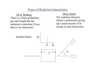
Types of Radiation Interactions. Many Small. All or Nothing. The radiation interacts almost continuously giving up a small amount of its energy at each interaction. There is a finite probability per unit length that the radiation is absorbed. If not, there is no interaction. Incident Beam. N.

E N D
Types of Radiation Interactions Many Small All or Nothing The radiation interacts almost continuously giving up a small amount of its energy at each interaction. There is a finite probability per unit length that the radiation is absorbed. If not, there is no interaction Incident Beam N E l
Types of Radiation Interactions Output beam The energy provides a marker for those photons of interest N N E E Attenuation tells us the depth. N N l l Angular spread of beam is maintained, thus well defined projection direction N N 0 0
Types of Interactions We Want y detector x Thus, the reduction in the beam intensity should be a property of the object along the line. Where is the linear attenuation coefficient and in general is a function of x and y -
Types of Interactions We Want Integrate along the path for a uniform material of length, x. In general, transmission thickness of absorber
Some details of photon interactions 1. “good” geometry - all photons that interact leave the measurement beam. 3 approaches 1) Restrict geometry to a narrow beam system. Collimator, place detector at infinity 2) Limit interaction to photo-electric (usually safe to assume that characteristic photons do not leave the sample) 3)Energy select detected photons Can define a build up factor to account for the additional photons at the detector or even in the sample itself.
Some details of photon interactions Consider a sample geometry with only a collimator at the output side Detector Source Collimator This volume element only sees the normal beam intensity . This volume element also sees the excess intensity from the buildup factor. So the buildup factor can contribute to the signal as well as the noise.
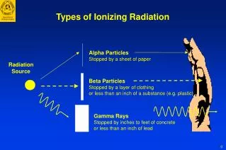
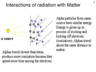
Attenuation Mechanisms (Simple Scatter) (a) Simple Scatter (Rayleigh Scattering) The incindent photon energy is much less than the binding energy of the electron in an atom. The photon is scattered without change of energy. Low energy relatively unimportant.
Attenuation Mechanisms (Photoelectric Effect) (b) Photoelectric effect The photon, slightly greater than gives up all of its energy to an inner shell electron, thereby ejecting it from the atom. The excited atom retains to the ground state with the emission of characteristic photons. Most of these are of relatively low energy and are absorbed by the material.
Attenuation Mechanisms (Compton Scattering) (c) Compton Scattering The photon energy is much greater than , and only part of this is given up during the interaction with an outer valence electron (the binding of valence electrons is relatively weak, hence the “free”). The photon is scattered with reduced energy and the energy of the electron is dissipated through ionizations.
Attenuation Mechanisms (Pair Production) (d) Pair Production A very high energy photon interacts with a nucleus to create an electron/positron pair. The mass of each particle is 9.11 x 10^-31 kg. So the minimum photon energy is: Both the electron and the positron lose energy via ionization until an anihilation event takes place yielding two photons of 0.51 MeV moving in oppoiste directions.
Ultrasound 1A 1mm 100mm 1cm 1m 100m X-ray Radio-frequency 1mm 100mm 1cm 1m 100m 100A damaging harmless C-H bond energy Tissue Transparency Windows of transparency in imaging via sound and electromagnetic radiation. The vertical scale measures absorption in tissue.
m dependence Mechanism E Z Energy Range in Soft Tissue simple scatter 1/E Z2 1-20 keV photoelectric 1/E3 Z3 1-30 keV Compton falls slowly with E independent 30 keV-20 MeV pair production rises slowly with E Z2 above 20 MeV Attenuation Mechanisms
Attenuation Mechanisms 2 Attenuation (log plot) total Compton Compton photoelectric pair simple scatter .01 .05 0.1 1 10 Photon energy (MeV) (log plot) .03 1.02 30 Attenuation mechanisms in water The optimum photon energy is about 30 keV (tube voltage 80-100 kV) where the photoelectric effect dominates. The Z3 dependence leads to good contrast: Zfat 5.9 Zmuscles 7.4 Zbone 13.9 Photoelectric attenuation from bone is about 11x that due to soft tissue, which is dominated by Compton scattering.
Beam Energy So, beam energy is important This does not include buildup factor or scattering but does include beam hardening
Beam Energy Also need to consider beam energy even if only photoelectric effect, since absorption rate depends on the energy. Thus, low energy photons deliver no useful information. N N E B Consider contrast agents, add a material to inhance contrast (more attenuation) k edge, minimal energy needed to have photoelectric effect with k shell electrons. 20 keV Increase the contrast, decrease the signal, increase the dose
Heterogeneous Case Interested in the heterogeneous case then Thus, in a continuosly varying medium a line integral over the sample and defined by the ray of interaction
Heterogeneous Case We wish to recontrast th linear attenuation coefficient . In 2D,
m/r (cm2/g) 5 2.5 2 1.0 BONE 0.5 MUSCLE 0.4 0.3 FAT 0.2 0.15 PHOTONENERGY (kev) 0.1 10 20 30 40 50 100 150 200 300 500 X-ray Attenuation Coefficients X-ray attenuation coefficients for muscle, fat, and bone, as a function of photon energy.
Unknown Electron ejected Characteristic Radiation Ionization event. Electron-electron interactions generates heat. This is the most common. Delta ray knocked out electron.
X-rays nucleus Bremsstrahlung - Breaking Radiation Coulombic interaction between electron and a nuclear charge For each interaction, the X-ray spectrum is white and the electron loses some energy. “True” Bremsstrahlung Spectrum Intensity Interaction
More Details On X-ray Tubes • electrons are boiled off filament • accelerated through a high vacuum from the cathode to the anode • electrons strike the anode, a tungsten target, and create X-rays • X-rays are emitted in all directions though only a cone is used • 99% of the electric energy is dissipated a heat into the anode. Typically less than 1% of the energy is converted into useful X-rays. • X-rays that are diverted into the target are absorbed and contribute to the production of heat.
Unknown But interactions filter out low energy Usually place some material between tube and object to further reduce low X-rays Need to take care in designing a filter so as not to create low energy charcteristic lines.
The X-Ray Spectrum (Changes in Voltage) The continuous spectrum is from electrons decelerating rapidly in the target and transferring their energy to single photons, Bremsstrahlung. The characteristic lines are a result of electrons ejecting orbital electrons from the innermost shells. When electrons from outer shells fall down to the level of the inner ejected electron, they emit a photon with an energy that is characteristic to the atomic transition.
The X-Ray Spectrum (Changes in Target Material) Increase in Z: Increase in X-ray intensity since greater mass and positive charge of the target nuclei increase the probability of X-ray emission total output intensity of Z Characteristic lines shift to higher energy, K and L electrons are more strongly held No change in