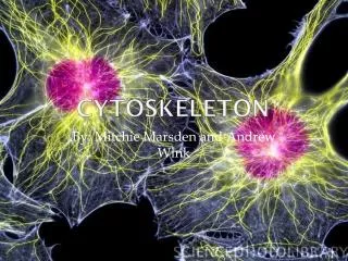
Cytoskeleton
A. Overview B. Experimental Methods C. Microtubules D. Microfilaments. Cytoskeleton. Overview Experimental Methods Microtubules Microfilaments. (Updated 4/9/08). A. Overview B. Experimental Methods C. Microtubules D. Microfilaments. A. Overview. Definition

Cytoskeleton
E N D
Presentation Transcript
A. Overview B. Experimental Methods C. Microtubules D. Microfilaments Cytoskeleton • Overview • Experimental Methods • Microtubules • Microfilaments (Updated 4/9/08)
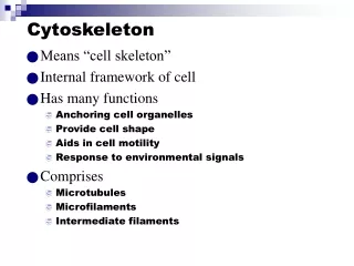
A. Overview B. Experimental Methods C. Microtubules D. Microfilaments A. Overview • Definition • Types of Cytoskeleton Fibers • Dynamic Polymerization/Depolymerization • Molecular Motors Alberts: Fig. 16 – 1, Panel 16 – 1, Panel 16 – 2, Fig. 16 –11, Fig 16 – 12, 16 – 8, 16 – 7, 16 – 10, 16 – 13, 16 – 14, 16 – 15, 16 – 16, 16 – 17, 16 – 19, 16 – 56, Table 16 – 1
A. Overview B. Experimental Methods C. Microtubules D. Microfilaments B. Experimental methods • Visualization Approaches • Light Microscopy • Fluorescence Microscopy • http://www.itg.uiuc.edu/exhibits/gallery/fluorescencegallery.htm • Digital/video Microscopy • Electron Microscopy • Genetic Approaches • Biochemical Approaches
A. Overview B. Experimental Methods C. Microtubules 1. Structure 2. MAPs 3. Functions 4. Microtubule Motors 5. MTOCs 6. Dynamic Properties 7. Flagella and Cilia D. Microfilaments C. Microtubules • Structure • Microtubule-associated proteins • Functions • Microtubule motors • Microtubule organizing centers • Dynamic properties of microtubules • Flagella and cilia
A. Overview B. Experimental Methods C. Microtubules 1. Structure 2. MAPs 3. Functions 4. Microtubule Motors 5. MTOCs 6. Dynamic Properties 7. Flagella and Cilia D. Microfilaments C.1. Microtubules: Structure • Structure • Alberts: Fig 16 – 11 • Structure and composition - hollow, tubular; found in most eukaryotic cells (cilia, spindle, flagella) • Outer diameter - 24 nm • Wall thickness - ~5 nm • May extend across cell length/breadth • Wall composed of globular proteins arranged in longitudinal rows (protofilaments) • Protofilaments are aligned parallel to tubule long axis • In cross section, consist of 13 protofilaments arrayed in circular pattern within wall
A. Overview B. Experimental Methods C. Microtubules 1. Structure 2. MAPs 3. Functions 4. Microtubule Motors 5. MTOCs 6. Dynamic Properties 7. Flagella and Cilia D. Microfilaments C.1. Microtubules: Structure • Each protofilament is assembled of dimeric building blocks (one a-tubulin & one b-tubulin; A heterodimer) organized in linear array along length of protofilament • Two types of tubulin subunits have similar 3D structure & fit tightly together • Protofilaments asymmetric (a-tubulin at one end, b-tubulin at other); All in single MT have same polarity; Each assembly unit has 2 nonidentical components (heterodimer) • All protofilaments of microtubule have same polarity; Thus so does full tubule (plus- & minus-end) • Plus end - fast-growing (b-tubulins on tip); Minus end - slow-growing (a-tubulins on tip)
A. Overview B. Experimental Methods C. Microtubules 1. Structure 2. MAPs 3. Functions 4. Microtubule Motors 5. MTOCs 6. Dynamic Properties 7. Flagella and Cilia D. Microfilaments C.2. Microtubules: MAPs • Microtubule-associated proteins • Alberts: Fig 16-40, 16-41 • MTs can assemble in vitro from purified tubulin, but MAPs are found with MTs isolated from cells; most found only in brain tissue; MAP4 has wider distribution • Have globular head domain that attaches to MT side & filamentous tail protruding from MT surface • May interconnect MTs to help form bundles (cross-bridges), increase MT stability, alter MT rigidity, influence MT assembly rate
A. Overview B. Experimental Methods C. Microtubules 1. Structure 2. MAPs 3. Functions 4. Microtubule Motors 5. MTOCs 6. Dynamic Properties 7. Flagella and Cilia D. Microfilaments C. 2. Microtubules: MAPs • MAP activity controlled by addition & removal of phosphate groups from particular amino acid residues by protein kinases & phosphatases, respectively; example - Alzheimer’s disease (AD) • Abnormally high MAP (tau) phosphorylation implicated in fatal neurodegenerative diseases like AD; neurofibrillary tangles in brains made of hyperphosphorylated tau; may help kill nerve cells • Excessively phosphorylated tau molecules are unable to bind to MTs; people with one of these diseases, a type of dementia called FTDP-17, carry mutations in tau gene, implicating it as cause
A. Overview B. Experimental Methods C. Microtubules 1. Structure 2. MAPs 3. Functions 4. Microtubule Motors 5. MTOCs 6. Dynamic Properties 7. Flagella and Cilia D. Microfilaments C. 3. Microtubules: Functions • Functions • Alberts: Table 16-2; Fig 16-23, 66 • Internal skeleton (scaffold) providing structural support & maintaining organelle position • Resist compression or bending forces on fiber; provide mechanical support like girders in `tall building; prevent distortion of cell by cytoplasmic contractions • MT distribution conforms to & helps determine cell shape: flattened, round cells - radiate from nuclear area; columnar epithelium - parallel to cell long axis; like aluminum rods support tent
A. Overview B. Experimental Methods C. Microtubules 1. Structure 2. MAPs 3. Functions 4. Microtubule Motors 5. MTOCs 6. Dynamic Properties 7. Flagella and Cilia D. Microfilaments C. 3. Microtubules: Functions • Elongated cell process (axon, axopods of heliozoan protists) - MTs oriented parallel to each other & axon or axopod long axis; help move things • In developing embryo, extend growing central NS axons to peripheral NS; inhibit (colchicine [CO], nocodazole [NO]) & outgrowth stops, regresses (collapses back to rounded cell body) • Found as core of axopodial processes of heliozoan protozoa; many MTs arranged in spiral with individual MTs traversing entire length of process
A. Overview B. Experimental Methods C. Microtubules 1. Structure 2. MAPs 3. Functions 4. Microtubule Motors 5. MTOCs 6. Dynamic Properties 7. Flagella and Cilia D. Microfilaments C. 3. Microtubules: Functions • Plants: play similar role in plants; affect shape indirectly by influencing cell wall formation; found in cortex just below membrane during interphase forming a distinct cortical zone
A. Overview B. Experimental Methods C. Microtubules 1. Structure 2. MAPs 3. Functions 4. Microtubule Motors 5. MTOCs 6. Dynamic Properties 7. Flagella and Cilia D. Microfilaments C. 3. Microtubules: Functions • Also have role in maintenance of cell internal organization (organelle placement) - disrupt MTs (CO, NO) —> Golgi disperses to cell periphery; goes back to cell center when inhibitors removed • Move macromolecules & organelles around cell in directed manner (intracellular motility) • Halt vesicle transport between compartments if disrupt MTs so transport dependent on them • Proteins made in neuron cell body move down axon (neurotransmitters, etc.) in vesicles
A. Overview B. Experimental Methods C. Microtubules 1. Structure 2. MAPs 3. Functions 4. Microtubule Motors 5. MTOCs 6. Dynamic Properties 7. Flagella and Cilia D. Microfilaments C. 3. Microtubules: Functions • Different materials move at different rates; fastest rate is 5 µm/sec (400 mm/day); vesicles seen attached to MTs • Structures & materials moving toward neuron terminals are said to move anterograde • Other structures, like endocytic vesicles that are formed at neuron terminals & carry regulatory factors from target cells, move from synapse to cell body in a retrograde direction • Ex.: axons filled with MTs, MFs & IFs; evidence suggests that both anterograde & retrograde movement are mediated mostly by MTs; video microscopy shows vesicles moving along MTs • Confirmed by EM of axons; molecular motors move vesicles along the MTs that serve as tracks
A. Overview B. Experimental Methods C. Microtubules 1. Structure 2. MAPs 3. Functions 4. Microtubule Motors 5. MTOCs 6. Dynamic Properties 7. Flagella and Cilia D. Microfilaments C. 3. Microtubules: Functions • Motile elements of cilia & flagella (more later) • Active components of mitotic/meiotic machinery; move chromosomes
A. Overview B. Experimental Methods C. Microtubules 1. Structure 2. MAPs 3. Functions 4. Microtubule Motors 5. MTOCs 6. Dynamic Properties 7. Flagella and Cilia D. Microfilaments C. 4. Microtubules: Motors • Microtubule motors • Alberts: Fig 16-58, 59, 60, 62, 63, 64, 67 • Motor proteins: convert chemical energy stored in ATP into mechanical energy that is used to move cellular cargo attached to motor • Types of cellular cargo transported by these molecular motors include: vesicles, organelles (mitochondria, lysosomes, chloroplasts), chromosomes, other cytoskeletal filaments • A single cell may contain dozens of different motor proteins, each specialized for moving a particular type of cargo in particular cell region
A. Overview B. Experimental Methods C. Microtubules 1. Structure 2. MAPs 3. Functions 4. Microtubule Motors 5. MTOCs 6. Dynamic Properties 7. Flagella and Cilia D. Microfilaments C. 4. Microtubules: Motors • Collectively, motor proteins are grouped into 3 broad families: myosins, kinesins, dyneins • Kinesins & dyneins move along MTs; myosins move along MFs; None known for ifs • Motor proteins move unidirectionally along their cytoskeletal track in a stepwise manner from one binding site to the next • As they move along, they undergo a series of conformational changes (a mechanical cycle) • Steps of mechanical cycle are coupled to chemical cycle, which provides energy fueling movement • Includes motor binding ATP, ATP hydrolysis, product (ADP & Pi) release & binding of new ATP • Binding & hydrolysis of 1 ATP moves motor a few nm along track; Cycles repeated many times
A. Overview B. Experimental Methods C. Microtubules 1. Structure 2. MAPs 3. Functions 4. Microtubule Motors 5. MTOCs 6. Dynamic Properties 7. Flagella and Cilia D. Microfilaments C. 4. Microtubules: Motors • Kinesins • Kinesins move vesicles/organelles from cell body to synaptic knobs; isolated in 1985 from squid giant axons; tetramer made of 2 identical heavy chains & 2 identical light chains; smallest & best understood • Large protein - pair of globular heads generate force by hydrolyzing ATP & bind MT; each head connected to a neck, a rodlike stalk & fan-shaped tail that binds cargo to be hauled • Diverse superfamily of kinesins - heads similar since roles similar; tails vary since they haul different cargoes
A. Overview B. Experimental Methods C. Microtubules 1. Structure 2. MAPs 3. Functions 4. Microtubule Motors 5. MTOCs 6. Dynamic Properties 7. Flagella and Cilia D. Microfilaments C. 4. Microtubules: Motors • In vitro mobility assay - kinesin-coated beads move to MT "+" end (axon tip); it is a "+" end-directed MT motor, therefore, kinesin responsible for anterograde movement • All MTs of axon are oriented with"-" ends facing cell body & "+" ends facing synaptic knobs • Moves through ATP-dependent cross-bridge cycle along single MT protofilament (rate proportional to [ATP]; up to ~1 µm/sec); at low concentrations, move slowly & see movement in distinct steps • Each step is ~8 nm in length, the spacing between tubulin dimers along protofilament • Appear to move 2 globular subunits (or 1 dimer at a time); usually toward membrane & "+" ends
A. Overview B. Experimental Methods C. Microtubules 1. Structure 2. MAPs 3. Functions 4. Microtubule Motors 5. MTOCs 6. Dynamic Properties 7. Flagella and Cilia D. Microfilaments C. 4. Microtubules: Motors • Kinesin possesses 2 motor domains that work by “hand-over-hand” mechanism; one always firmly attached to MT • 2 heads of kinesin behave in coordinated manner, so that they are always present at different stages in their chemical & mechanical cycles at a given time • When one head binds to MT, the interaction induces a conformational change in adjacent neck region of motor protein; it swings the other head forward toward binding site on next dimer • Force generated by head catalytic activity leads to swinging movement of neck • A kinesin molecule walks along a MT, hydrolyzing one ATP with each step
A. Overview B. Experimental Methods C. Microtubules 1. Structure 2. MAPs 3. Functions 4. Microtubule Motors 5. MTOCs 6. Dynamic Properties 7. Flagella and Cilia D. Microfilaments C. 4. Microtubules: Motors • Conventional kinesin (discovered in 1985) is only one member of a superfamily of related kinesins • Mammalian genome sequence analysis leads to estimate that mammals make >50 different kinesins • Heads of kinesins have related amino acid sequences, reflecting common evolutionary ancestry & their similar role in moving along MTs • In contrast, kinesin tails have diverse sequences, reflecting variety of cargo different proteins haul
A. Overview B. Experimental Methods C. Microtubules 1. Structure 2. MAPs 3. Functions 4. Microtubule Motors 5. MTOCs 6. Dynamic Properties 7. Flagella and Cilia D. Microfilaments C. 4. Microtubules: Motors • Most kinesins travel toward the "+" end; but one small subfamily of kinesins (including the Drosophila Ncd protein) moves toward the MT "-" end • one would expect that the heads of "+"- & "-"-directed would have a different structure since the heads contain the catalytic core of the motor domain • But the heads are virtually indistinguishable; instead differences in direction of movement are determined by differences in the adjacent neck regions of the two proteins • When the head of a "-" end-directed Ncd molecule is joined to the neck-stalk portions of a kinesin molecule, the hybrid protein moves toward the "+" end of a MT track • Even if the hybrid has a catalytic domain that would normally move toward the "-" end of a MT, as long as it is joined to the neck of a "+" end motor, it moves in the "+" direction
A. Overview B. Experimental Methods C. Microtubules 1. Structure 2. MAPs 3. Functions 4. Microtubule Motors 5. MTOCs 6. Dynamic Properties 7. Flagella and Cilia D. Microfilaments C. 4. Microtubules: Motors • A third subfamily of kinesinlike proteins is incapable of movement: kinesins of this group, like KXKCM1, are thought to destabilize MTs rather than acting as MT motors
A. Overview B. Experimental Methods C. Microtubules 1. Structure 2. MAPs 3. Functions 4. Microtubule Motors 5. MTOCs 6. Dynamic Properties 7. Flagella and Cilia D. Microfilaments C. 4. Microtubules: Motors • Cytoplasmic Dyneins • Dyneins - first MT-associated motor found (1963); responsible for moving cilia & flagella • Thought to be ubiquitous eukaryotic motor protein; related protein found in variety of nonneural cells • Cilia/flagella form of protein was called axonemal dynein; its new relatives were called cytoplasmic dynein • Huge protein (~1.5 million daltons); 2 identical heavy chains & variety of intermediate & light chains • Each dynein heavy chain forms large globular head (~10X larger than a kinesin head) that generates force; moves along MT toward "-" end
A. Overview B. Experimental Methods C. Microtubules 1. Structure 2. MAPs 3. Functions 4. Microtubule Motors 5. MTOCs 6. Dynamic Properties 7. Flagella and Cilia D. Microfilaments C. 4. Microtubules: Motors • Suggested roles of cytoplasmic dynein • Force generating agent for chromosome movement in mitosis • "-"-directed MT motor for Golgi complex positioning & movement of vesicles/organelles through cytoplasm • In nerve cells, cytoplasmic dynein involved in axonal retrograde organelle movement (toward cell body & cell center) & anterograde movement of MTs • Fibroblasts & other nonneural cells: may move varied membranous organelles (endosomes, lysosomes, ER-derived vesicles going toward Golgi) from periphery toward cell center
A. Overview B. Experimental Methods C. Microtubules 1. Structure 2. MAPs 3. Functions 4. Microtubule Motors 5. MTOCs 6. Dynamic Properties 7. Flagella and Cilia D. Microfilaments C. 4. Microtubules: Motors • Cytoplasmic dynein does not interact directly with membrane-bounded cargo, but requires intervening multisubunit complex, dynactin that may regulate dynein activity & help bind it to MT • Present model may be overly simplistic: kinesin & cytoplasmic dynein move similar materials in opposite directions over the same railway network • Individual organelles may bind kinesin & dynein simultaneously although only one is active at given time; myosin may also be present on some of these organelles
A. Overview B. Experimental Methods C. Microtubules 1. Structure 2. MAPs 3. Functions 4. Microtubule Motors 5. MTOCs 6. Dynamic Properties 7. Flagella and Cilia D. Microfilaments C. 5. Microtubules: MTOCS • Microtubule-organizing centers • Alberts: Panel 16-1; Fig 16-29, 30, 31, 32, 33 Function of MT in living cell depends on its location & orientation, thus it is important to understand why a MT assembles in one place as opposed to another • controlled by MT-organizing centers (MTOCs)
A. Overview B. Experimental Methods C. Microtubules 1. Structure 2. MAPs 3. Functions 4. Microtubule Motors 5. MTOCs 6. Dynamic Properties 7. Flagella and Cilia D. Microfilaments C. 5. Microtubules : MTOCS • Assembly of MTs from ab-dimers occurs in 2 distinct phases • Nucleation - slower; small portion of MT initially formed; occurs in association with specialized structures in vivo called microtubule-organizing centers (MTOCs); centrosome is example • Elongation - more rapid
A. Overview B. Experimental Methods C. Microtubules 1. Structure 2. MAPs 3. Functions 4. Microtubule Motors 5. MTOCs 6. Dynamic Properties 7. Flagella and Cilia D. Microfilaments C. 5. Microtubules : MTOCS • Centrosomes - complex structure with 2 barrel-shaped centrioles surrounded by amorphous, electron dense pericentriolar material (PCM) • In animal cells, cytoskeleton MTs typically form in association with centrosome • Centrioles: cylindrical; ~0.2 nm dia & typically ~twice as long; usually with 9 evenly spaced fibrils • Each fibril seen in cross section to be composed of 3 fused MTs (A, [the only complete one]; B & C), A is attached to centriole center by radial spoke
A. Overview B. Experimental Methods C. Microtubules 1. Structure 2. MAPs 3. Functions 4. Microtubule Motors 5. MTOCs 6. Dynamic Properties 7. Flagella and Cilia D. Microfilaments C. 5. Microtubules : MTOCS • 3 MTs of each triplet arranged in pattern that gives centriole a characteristic pinwheel appearance • Centrioles usually in pairs at right angles to each other near cell center just outside nucleus • Extraction of isolated centrosomes with 1 M potassium iodide removes ~90% of PCM protein leaving behind spaghetti-like scaffold of insoluble fibers • Centrosomes are sites of convergence of large numbers of MTs
A. Overview B. Experimental Methods C. Microtubules 1. Structure 2. MAPs 3. Functions 4. Microtubule Motors 5. MTOCs 6. Dynamic Properties 7. Flagella and Cilia D. Microfilaments C. 5. Microtubules : MTOCS • MT polymerization & disassembly - treat with poisons (CO, NO) or cold —> MTs disassemble; much has been learned about their disassembly & reassembly in cultured animal cells in this way • Observe assembly when cells warmed or poisons removed; fix at various times after & stain with fluorescent anti-tubulin ABs • Within a few minutes of inhibition removal, 1 or 2 bright fluorescent spots seen in cytoplasm • Within 15 - 30 minutes, number of labeled filaments radiating from these foci rises dramatically
A. Overview B. Experimental Methods C. Microtubules 1. Structure 2. MAPs 3. Functions 4. Microtubule Motors 5. MTOCs 6. Dynamic Properties 7. Flagella and Cilia D. Microfilaments C. 5. Microtubules : MTOCS • In EM: MTs radiate out from centrosome; MTs don't actually penetrate into centrosome & contact centrioles, but terminate in dense pericentriolar material residing at centrosome periphery • PCM apparently initiates MT formation; centrioles not involved in MT nucleation, but they probably play a role in recruiting surrounding PCM during centrosome assembly
A. Overview B. Experimental Methods C. Microtubules 1. Structure 2. MAPs 3. Functions 4. Microtubule Motors 5. MTOCs 6. Dynamic Properties 7. Flagella and Cilia D. Microfilaments C. 5. Microtubules : MTOCS • Centrosome typically situated near center of cell, just outside nucleus • Columnar epithelium - centrosome moves from cell center to apical region just beneath cortex; cytoskeletal MTs emanate from site, extending toward nucleus & basal surface of cell • Regardless of location, centrosomes are sites of MT nucleation; polarity is always the same: "-" end at centrosome, "+" (growing) end at opposite tip • Thus, even though MTs are nucleated at MTOC, they are elongated at opposite end of polymer
A. Overview B. Experimental Methods C. Microtubules 1. Structure 2. MAPs 3. Functions 4. Microtubule Motors 5. MTOCs 6. Dynamic Properties 7. Flagella and Cilia D. Microfilaments C. 5. Microtubules : MTOCS • Not all MTs associated with centrosome; • some animal cells (mouse oocytes) lack centrosomes entirely, but still make spindle • MTs of axon are not associated with centrosome, which is located in cell body, but they may be formed at centrosome, then released from that MTOC & carried to axon by motor proteins
A. Overview B. Experimental Methods C. Microtubules 1. Structure 2. MAPs 3. Functions 4. Microtubule Motors 5. MTOCs 6. Dynamic Properties 7. Flagella and Cilia D. Microfilaments C. 5. Microtubules : MTOCS • Basal bodies & other MTOCs • Centrosomes are not the only MTOCs in cells; basal bodies at base of cilia & flagella serve as origin of ciliary & flagellar MTs; MTs grow out of them • Basal body cross-section looks like centriole; in fact, the two can give rise to one another • Sperm flagellum arises from basal body derived from sperm centriole that had been part of meiotic spindle of spermatocyte from which the sperm arose • Conversely, sperm basal body typically becomes centriole during fertilized egg's first mitotic division of fertilized egg
A. Overview B. Experimental Methods C. Microtubules 1. Structure 2. MAPs 3. Functions 4. Microtubule Motors 5. MTOCs 6. Dynamic Properties 7. Flagella and Cilia D. Microfilaments C. 5. Microtubules : MTOCS • Plant MTOC - lack both centrioles & centrosomes; MTOCs more dispersed than those of animals • In plant endosperm cells, the primary MTOC consists of patches of material situated at outer surface of nuclear envelope from which cytoskeletal MTs emerge • MT nucleation also thought to occur throughout plant cell cortex
A. Overview B. Experimental Methods C. Microtubules 1. Structure 2. MAPs 3. Functions 4. Microtubule Motors 5. MTOCs 6. Dynamic Properties 7. Flagella and Cilia D. Microfilaments C. 5. Microtubules : MTOCS • MT nucleation • Despite diverse appearances, all MTOCs play similar roles in all cells • Control number of MTs that form & their polarity • Control the number of protofilaments that make up their walls • Control the time & location of MT assembly
A. Overview B. Experimental Methods C. Microtubules 1. Structure 2. MAPs 3. Functions 4. Microtubule Motors 5. MTOCs 6. Dynamic Properties 7. Flagella and Cilia D. Microfilaments C. 5. Microtubules : MTOCS • All MTOCs share a common protein component, g-tubulin (discovered in mid-1980s); it is ~0.005% of total cell protein while a- & b-tubulins are 2.5% of total nonneural cell protein • Fluorescent anti-g-tubulin antibodies (ABs) stain all MTOCs, like centrosome PCM; suggests it is critical component in MT assembly & nucleation • Microinject anti-g-tubulin AB into living cell —> blocks MT reassembly after depolymerization by drugs or cold temperatures • Genetically engineered fungi lacking functional g-tubulin gene cannot assemble normal MTs
A. Overview B. Experimental Methods C. Microtubules 1. Structure 2. MAPs 3. Functions 4. Microtubule Motors 5. MTOCs 6. Dynamic Properties 7. Flagella and Cilia D. Microfilaments C. 5. Microtubules : MTOCS • Nucleation mechanism revealed by structure/composition studies of PCM at centrosome periphery • Insoluble fibers of PCM are thought to serve as attachment sites for ring-shaped structures that have same diameter as MTs (25 nm) & contain g-tubulin • Ring-shaped structures found when centrosomes were purified & incubated with gold-labeled anti-g-tubulin ABs —> cluster in rings/semi-circles at MT minus ends (ends embedded in PCM) • Isolate similar ring-shaped complexes (g-TuRCs) from cell extracts; nucleate MT assembly in vitro
A. Overview B. Experimental Methods C. Microtubules 1. Structure 2. MAPs 3. Functions 4. Microtubule Motors 5. MTOCs 6. Dynamic Properties 7. Flagella and Cilia D. Microfilaments C. 5. Microtubules : MTOCS • Model - helical array of 13 g-tubulin subunits forms open, ring-shaped template on which first row of ab-tubulin dimers assemble; • Only a-tubulin of heterodimer can bind to ring of g-subunits, establishing polarity of entire MT • 2 other tubulin isoforms d-tubulin & e-tubulin have also been identified in centrosomes, but their function has not been determined
A. Overview B. Experimental Methods C. Microtubules 1. Structure 2. MAPs 3. Functions 4. Microtubule Motors 5. MTOCs 6. Dynamic Properties 7. Flagella and Cilia D. Microfilaments C. 6. Microtubules : Dynamic • Dynamic properties of microtubules • Alberts: Table 16-2; Fig 16-16, 16-17 • MTs vary markedly in stability even though similar morphologically - spindle/cytoskeleton labile; mature neuron MTs less labile; cilia/flagella very stable; lability allows cell to respond to stimuli • Cilia/flagella MTs are stabilized by MAP attachment & by enzymatic modification (e. g. acetylation) of specific amino acid residues within tubulin subunits • Labile MTs in living cells can be disassembled without disrupting other cell structures via a number of treatments
A. Overview B. Experimental Methods C. Microtubules 1. Structure 2. MAPs 3. Functions 4. Microtubule Motors 5. MTOCs 6. Dynamic Properties 7. Flagella and Cilia D. Microfilaments C. 6. Microtubules : Dynamic • Treatments that cause MT disassembly; usually interfere with noncovalent bonds holding them together • Cold temperatures • Hydrostatic pressure • Elevated Ca2+ concentration • Variety of chemicals (often used in chemotherapy) - CO, vinblastine, vincristine, NO, podophyllotoxin
A. Overview B. Experimental Methods C. Microtubules 1. Structure 2. MAPs 3. Functions 4. Microtubule Motors 5. MTOCs 6. Dynamic Properties 7. Flagella and Cilia D. Microfilaments C. 6. Microtubules : Dynamic • Some treatments (taxol) disrupt MT dynamic activity & act by doing the opposite; inhibit disassembly • Taxol binds MT polymer & thus prevents disassembly; cell cannot build new MT structures
A. Overview B. Experimental Methods C. Microtubules 1. Structure 2. MAPs 3. Functions 4. Microtubule Motors 5. MTOCs 6. Dynamic Properties 7. Flagella and Cilia D. Microfilaments C. 6. Microtubules : Dynamic • Cytoskeletal MT lability reflects fact that they are polymers formed by noncovalent association of dimers; subject to depolymerization/repolymerization as cell needs change • Dramatic changes in MT spatial organization may be achieved by combination of 2 separate mechanisms • Rearrangement of existing MTs • Disassembly of existing MTs & reassembly of new ones in different cell regions
A. Overview B. Experimental Methods C. Microtubules 1. Structure 2. MAPs 3. Functions 4. Microtubule Motors 5. MTOCs 6. Dynamic Properties 7. Flagella and Cilia D. Microfilaments C. 6. Microtubules : Dynamic • Study of MT dynamics in vitro - suggest that cytoskeleton can rapidly remodel & respond to stimuli • Early studies established that GTP binding to b -subunit required for MT assembly; GTP hydrolysis not needed for binding, but it is hydrolyzed soon after dimer attached to MT end; GDP stays bound • After dimer is released from MT during disassembly & enters soluble pool, GDP is replaced by GTP, thus recharging dimer so that it can add to polymer again • A GTP molecule is also bound to a-tubulin subunit, but it is not exchangeable & it is not hydrolyzed after subunit incorporation
A. Overview B. Experimental Methods C. Microtubules 1. Structure 2. MAPs 3. Functions 4. Microtubule Motors 5. MTOCs 6. Dynamic Properties 7. Flagella and Cilia D. Microfilaments C. 6. Microtubules : Dynamic • Assembly is not energetically inexpensive since it includes GTP hydrolysis, but it does allow the cell to control assembly & disassembly independently • A dimer being added to MT has a bound GTP; dimer being released from MT has bound GDP • GDP- & GTP dimers have different conformations & participate in different reactions; the ends of growing & shrinking MTs have different structures
A. Overview B. Experimental Methods C. Microtubules 1. Structure 2. MAPs 3. Functions 4. Microtubule Motors 5. MTOCs 6. Dynamic Properties 7. Flagella and Cilia D. Microfilaments C. 6. Microtubules : Dynamic • The above facts lead to the following model: • When a MT is growing, the "+" end is present as an open sheet to which GTP-dimers are added • During rapid growth periods, tubulin dimers are added faster than GTP can be hydrolyzed • The resultant cap of GTP-dimers on MT at protofilament ends is thought to favor the addition of more subunits & hence MT growth • However, MTs with open ends thought to undergo spontaneous reaction leading to tube closure
A. Overview B. Experimental Methods C. Microtubules 1. Structure 2. MAPs 3. Functions 4. Microtubule Motors 5. MTOCs 6. Dynamic Properties 7. Flagella and Cilia D. Microfilaments C. 6. Microtubules : Dynamic • tube closure is accompanied by hydrolysis of bound GTP, changing tubulin dimer conformation —> resultant mechanical strain destabilizes MTs • Strain is released as protofilaments curl out from tubule & catastrophically depolymerize • Disassembly can occur remarkably fast, especially in vivo, which allows very rapid MT cytoskeleton disassembly
A. Overview B. Experimental Methods C. Microtubules 1. Structure 2. MAPs 3. Functions 4. Microtubule Motors 5. MTOCs 6. Dynamic Properties 7. Flagella and Cilia D. Microfilaments C. 6. Microtubules : Dynamic • Study of MT dynamics in vivo: dynamic character of MT cytoskeleton inside cell is best revealed by microinjecting labeled tubulin into nondividing cultured cell • Inject labeled tubulin into nondividing cultured cell —> labeled subunits rapidly incorporated into preexisting cytoskeleton MTs, even in absence of any obvious morphological change • Watch cell with fluorescent-labeled MTs over time —> some MTs grow, others shrink; dynamic • Both growth & shrinkage in vivo occur predominantly at "+" end of polymer, the end located opposite the centrosome (or other MTOC)
A. Overview B. Experimental Methods C. Microtubules 1. Structure 2. MAPs 3. Functions 4. Microtubule Motors 5. MTOCs 6. Dynamic Properties 7. Flagella and Cilia D. Microfilaments C. 6. Microtubules : Dynamic • Single MTs switch randomly & unpredictably between growing & shrinking (dynamic instability) • MTs shrink faster than they grow, so in a matter of minutes, MTs disappear & are replaced by new MTs that grow out from centrosome
A. Overview B. Experimental Methods C. Microtubules 1. Structure 2. MAPs 3. Functions 4. Microtubule Motors 5. MTOCs 6. Dynamic Properties 7. Flagella and Cilia D. Microfilaments C. 7. Microtubules: Flagella • Cilia and flagella structure • Alberts: Fig 16-80, 81, 82, 83, 84 • Entire ciliary or flagellar projection is covered by membrane continuous with cell membrane • Cilium core (axoneme) contains an array of MTs that run longitudinally through entire organelle • Usually 9 peripheral doublet MTs surrounding central pair of single MTs; known as 9 + 2 pattern or array; all MTs in array have same polarity ("+" ends at tip, "-" ends at base) • Doublets - 1 complete (A tubule; 13 subunits) MT; 1 incomplete (B tubule) MT with 10 or 11 subunits