Download
1 / 20
200 likes | 383 Views
Antibody Lab B Cell Ligand Screen. Quantify ligand-induced changes in site-specific phosphorylation of selected signaling proteins. Erica Turon, technician and Heping Han, Lead Scientist. Antibody Lab B Cell Ligand Screen. Aim : sample diversity of responses to inputs

E N D
Antibody LabB Cell Ligand Screen Quantify ligand-induced changes in site-specific phosphorylation of selected signaling proteins Erica Turon, technician and Heping Han, Lead Scientist
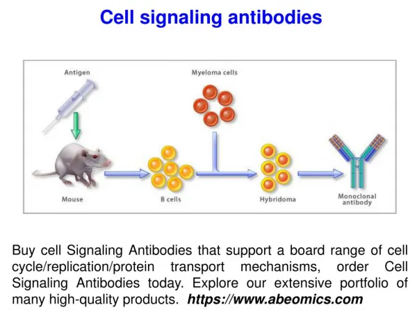
Antibody LabB Cell Ligand Screen • Aim: sample diversity of responses to inputs • Approach: Probe Western blots with mixture of site specific, phosphorylation sensitive antibodies • Goal: resolve and quantify 5-7 phosphoproteins per lane of gel • Progress: 2 mixtures of antibodies for 11 phosphoproteins
Mix 1 Stat 6: Y641 p90RSK: S381 Akt: S473 ERK 1 T202/Y204 ERK 2 T183/Y185 Mix 4 PKCm S916 Stat 3 Y705 NFkB p65 S536 JNK long T183/Y185 JNK short T183/Y185 p38 MAPK T180/Y182 Targets of Current Mixes of Phosphospecific Antibodies Criteria: single band of appropriate size, ligand sensitive Tested 64 different antibody preparations (1 out of 7) All antibodies from Cell Signaling Technology, except one
B Cell Receptor Map * *
Phosphospecific Antibody Mix 1 p90RSK Stat6 Pos.Cont.A Untreated IL-4 Anti-IgM ERKs Akt Anti-IgM = 0.3 mM, IL-4 = 0.4 nM, Positive control A = Anti-IgM + IL-4 All treatment was 5 min
Ligand Screen Time Courses + Control Untreated Untreated + Control <Stat6 <p90RSK <Akt >ERKs <Gb EWC020314B_EA Cell ID = EWC020319D Anti-IgM = 0.3 mM; IL-4 = 0.34 nM ; CD-40 = 65 nM
Imaging • No film or radioactivity • ECL Plus (has fluorescent by-product) • Imager detects fluorescence • ImageQuant software to save image Biren Zhao with Storm 860 Imager from Molecular Dynamics
Quantification • Nick Wong: searched for better software, implemented • ImageMaster: Automatically finds lanes, semi-automatic for bands
Interleukin 4 Phosphorylation of Stat 6 • 50 fold increase in phosphorylation of Stat6 • Single scale: Cannot see changes in phosphorylation of other proteins
Interleukin 4 • Two y axes scaled differently • Reveals phosphorylation of Akt (fuscia)
Additional Ligands:Gi-mediated Responses • Sphingosine 1-phosphate and Chemokines: SDF, BLC, ELC, SLC • Increased phosphorylation of Akt, ERKs and p90RSK with • max. response at first time point • More rapid initial decline of ERKs • Time course pattern similar to B cell receptor • Gq, Gs pathways: no effect of terbutaline, PGE2, LPA, 2-MA
Antibody Mix 1Additional Ligands with No Response IL-10 Interferon g IGF-1 TNF a Leukotriene B4 fMLP Neurokinin B Responses with Antibody Mix 4?
Phosphospecific Antibody Mix 4 PKCm Stat3 Pos. Cont. B NF kB p65 JNK p38MAPK EA239_April 1002 Positive Control B = 0.34 nM IL-10 and 65 nM anti-CD40 treated B cells for 5 min.
CD40 Signaling Map * * *
Phosphoprotein Antibody Mix 4Responses to Anti-CD40 • Early response: phosphorylation of NFkB p65 • JNKs: • delayed response (similar to ERKs and Akt) • separate y axis
Phosphoprotein Antibody Mix 4Responses to IL-4 and Anti-IgM • IL-4: Rate of Stat 3 phosphorylation slower than Stat 6 • Anti-IgM: modest responses
Phosphoprotein Antibody Mix 4Ligand Screen • Anti-IgM, Anti-CD40 and IL-4 quantified • n=2 for data shown • Data on website soon Becky Fulin, Technician
Our Group Antibody Lab: Lead scientist: Heping Han Technicians: Eduardo Arteaga, Becky Fulin, Nick Wong, Biren Zhao Dallas Labs: Data Manager: Lonnie Sorrells Administrator: Kim Edwards
Future Goals Eduardo Arteaga • Ligand Screens • B cell • WEHI 231 • Myocytes • Multi-ligand • Luminex fluorescent bead technology • Determine concentration of signaling proteins • Focus on PIP3 Modules in WEHI 231 cells • Characterize/utilize phosphospecific antibodies • Collaboration with Cell Signaling Technology