Slide Note
0 likes | 17 Views
A peripheral blood smear, also known as a differential leukocyte count (DLC), is a procedure where a thin layer of blood is examined under a microscope to identify and count different types of blood cells. This test helps in detecting infections, monitoring blood diseases like leukemia, and diagnosing hematological problems. The DLC focuses on three main types of white blood cells: granulocytes (neutrophils, eosinophils, basophils) and agranulocytes (lymphocytes, monocytes). By preparing and staining a blood smear, healthcare professionals can assess the various types of white blood cells to aid in diagnosing conditions. Leishman's stain is commonly used for this purpose, as it allows for clear visualization of different cell types.

E N D
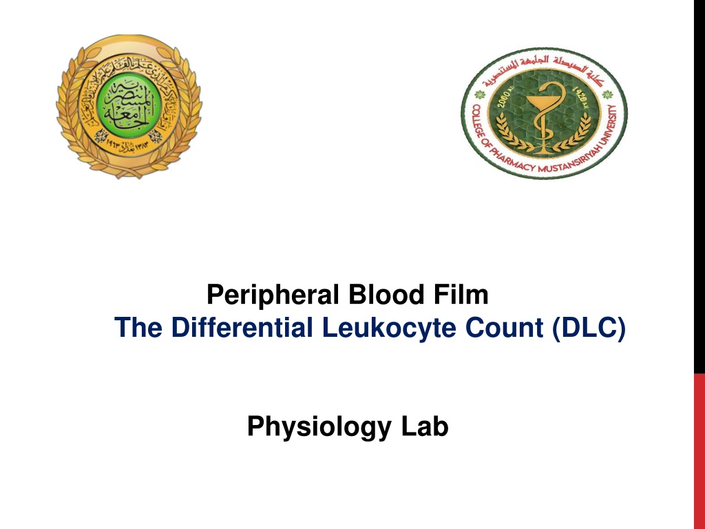
Peripheral Blood Film The Differential Leukocyte Count (DLC) Physiology Lab
peripheral blood film the differential leukocyte count (DLC) Definition: Blood film or peripheral blood smear is a thin layer of blood smeared on a slide and then stained in such a way to allow the various blood cells to be examined microscopically. A blood smear is a blood test that gives information about the number and shape of blood cells. The cell types are examined investigate hematological problems (disorders of the blood) Goals: • The student will prepare a blood smear which is even, smooth and have an acceptable feathered edge. • To become familiar with the various types of white blood cells under a microscope to
APPLICATIONS OF BLOOD SMEAR: • to detect infection or inflammation, determine the effects of possible poisoning by chemicals, drugs, chemotherapy, radiation, etc. • DLC is also done to monitor blood diseases like leukemia, or to detect allergic and parasitic infections. • The determination of each type of WBC helps in diagnosing the condition because a particular type may show an increase or decrease.
BACKGROUND • There are three main cells within the blood that the test focuses on: red cells: (which carry oxygen throughout the body) white cells: (which function as part of the body’s immune system) Platelets: (which are important for blood clotting).
TYPES OF WBCS Granulocytes are larger than RBCs and have lobed nuclei and granules in their cytoplasm. • Neutrophils • Eosinophils • Basophils
GRANULOCYTES 1-Neutrophils: 40-70% of WBCs, 3-7 lobed nucleus, pale lilac or pink cytoplasm contains very fine granules which are difficult to see. They are active phagocytes and fight bacterial invasions (important in inflammatory response) as well as cleaning up debris.
GRANULOCYTES 2-Eosinophils: 1-4% of WBCs, bilobed nucleus, large red/orange cytoplasmic granules. Important in ending allergic reactions (phagocytize antibody-bound allergens) and fighting parasitic worms.
GRANULOCYTES 3- Basophils: less than 1% of WBCs, nucleus often U or S shaped with indentations, large dark purple cytoplasmic granules. Mediate inflammatory response (release histamine and other molecules) during allergic responses and parasitic infections.
TYPES OF WBCS Agranulocytes have no observable granules and nuclei are usually roughly spherical. Monocytes Lymphocytes
AGRANULOCYTES 1-Lymphocytes: 20-40% of WBCs, about the size of a RBC, dark blue or purple nucleus, sparse gray/blue cytoplasm. Important role in immune system.
AGRANULOCYTES 2- Monocytes: 4-8% of WBCs, largest of the WBCs, dark blue nucleus, abundant gray/blue cytoplasm.They are active phagocytes and considered important in long-term clean up.
MATERIALS AND INSTRUMENTS 1-Whole blood using EDTA as anticoagulant or capillary blood drawn from a finger or toe puncture. 2- Glass slide 3- Microscope 4-Alcohol 70% 5- Lancet 6- Leishman's stain Leishman’s stain: It is probably one of the simplest and most precise methods of staining blood for diagnostic purposes. It contains a compound dye—eosinate of methylene-blue dissolved in acetone-free methyl alcohol.
Eosin. It is an acidic dye (negatively charged) and stains basic (positive) particles— granules of eosinophils, and RBCs a pink color. Methylene-blue. It is a basic dye (positively charged) and stains acidic (negatively charged) granules in the cytoplasm, nuclei of leukocytes, especially the granules of basophils, a blue-violet color. Acetone-free and water-free absolute methyl alcohol. The methyl alcohol is a fixative and must be free from acetone and water. It serves two functions: a. It fixes the blood smear to the glass slide. The alcohol precipitates the plasma proteins, which then act as a ‘glue’which attaches (fixes) the blood cells to the slide so that they are not washed away during staining. b. The alcohol preserves the morphology and chemical status of the cells.
PREPARATION OF BLOOD SMEAR Two types of smear can be prepared: a. Thin blood smear – for demonstration and differentiation of leukocytes. 1. Take a clean slide. Obtain capillary blood directly from the finger or EDTA anti-coagulated blood.Add one small drop of blood to one end. 2. 3. Take another clean slide, and holding at an angle of about 45°. Touch the blood with one end of the slide so the blood runs along the edge of the slide by capillary action. Push carefully along the length of the first slide to produce a thin smear of blood. 4. Make 2 smears, allow to air dry, and label clearly. Once dried place in the provided slide transport containers
b. Thick blood smear – for diagnosis of blood protozoan parasites and blood abnormalities, ex- anemia. 1. Place a small drop of blood in the center of the pre-cleaned, labeled slide. 2. Using the corner of another slide or an applicator stick, spread the droA thick smear of proper density is one which, if placed (wet) over newsprint, allows you to barely read the words. 3. Lay the slides flat and allow the smears to dry thoroughly (protect from dust and insects!). p in a circular pattern until it is the size of a dime (1.5 cm2).
IDENTIFYING A LEUKOCYTE • A leukocyte is identified from its size, its nucleus, and the cytoplasm—its color, whether vesicles (granules) are visible or not, their color and size if visible, and the cytoplasm/nucleus ratio.
CALCULATION • Count each WBC seen and record on a differential cell counter until 100 WBCs have been counted. For instance if 25 of the 100 WBCs where lymphocytes, then the percentage of lymphocytes is 25%. • The normal range percentage of the different types of WBCs is as follows: Neutrophils 50-70% Eosinophils 1-4% Basophils 0.4% Monocytes 2-8% Lymphocytes 20-40%
Worksheet Lab 6 Names of the students: Date---------- Group-------- Name of the experiment: Aim of the experiment: Materials Procedure: Prepare the blood smear Clean two slide, one to be covered with the blood film and one to be used as spreader. Clean the finger with alcohol, allow it to dry and then prick it with a disposable lancet to obtain a drop of blood. Make a fine touch of one end of a slide with the drop of blood (only a small amount is required). Place the edge of the other slide on the surface of the first slide just in front of the drop of blood and at an angle of 45˚. Draw the spreader back until it makes contact with the drop of blood. Push the spreader slowly and smoothly to the other end of the slide. Allow the film to dry at room temperature Staining the blood smear by:- Put the dried slide on a staining rack. Carefully drop Leishman's stain onto the blood film until the film is covered. Allow the stain to act for one to two minutes. Add distilled water to the stain, this gives dilution of 1:1 or 1:2 The diluted stain should act for 15-30 minutes Then wash it off with distilled water, continue washing until the film has a pink color. Shake off excess water and allow it to dry at room temperature. Examine the prepared slide under a microscope Results Discussion:


![[PDF] Free Download The Blood Mirror By Brent Weeks](https://cdn4.slideserve.com/8010772/slide1-dt.jpg)
![[PDF] Free Download Blood Of Iax By Robbie MacNiven](https://cdn4.slideserve.com/8018502/slide1-dt.jpg)
![[PDF] Blood Communion](https://cdn4.slideserve.com/8031337/slide1-dt.jpg)
![[PDF] Free Download Fire and Blood By George R.R. Martin & Doug Wheatley](https://cdn4.slideserve.com/8142107/slide1-dt.jpg)