Download
1 / 43
430 likes | 609 Views
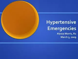
1000 hypertensive patients. 940 essential hypertension. 60 secondary hypertension. 50 renal hypertension 5 reno-vascular hypertension 5 endocrine hypertension. 2 pheochromocytomas 2 primary hyperaldosteronism 1 other endocrine cause. avant 2000.

E N D
1000 hypertensive patients 940 essential hypertension 60 secondary hypertension 50 renal hypertension 5 reno-vascular hypertension 5 endocrine hypertension 2 pheochromocytomas 2 primary hyperaldosteronism 1 other endocrine cause avant 2000
Pourquoi s’intéresser aux causes endocriniennes d’hypertension ? • Les mécanismes étiopathogéniques d’ hypertension endocrinienne sont aussi impliqués dans l’hypertension essentielle • activation de l’axe rénine-angiotensine-aldostérone • hyperactivité du système adréno-sympathique • L’hypertension endocrinienne est sous-diagnostiquée ! • hyperaldostéronisme primaire fréquent • bcp de phéochromocytomes restent longtemps occultes • incidentalome et syndrome de Cushing infraclinique
15 % HTA Adrenal incidentaloma multicentric italian study ( Mantero et al, JCEM, 2000 ) : - 1004 incidentalomas + hormonal work-up - malignant and symptomatic tumors excluded • non-secreting tumor 85 % • subclinical Cushing’s syndrome (SCS) 9,2 % • “silent” pheochromocytoma 4,2 % • primary hyperaldosteronism 1,6 %
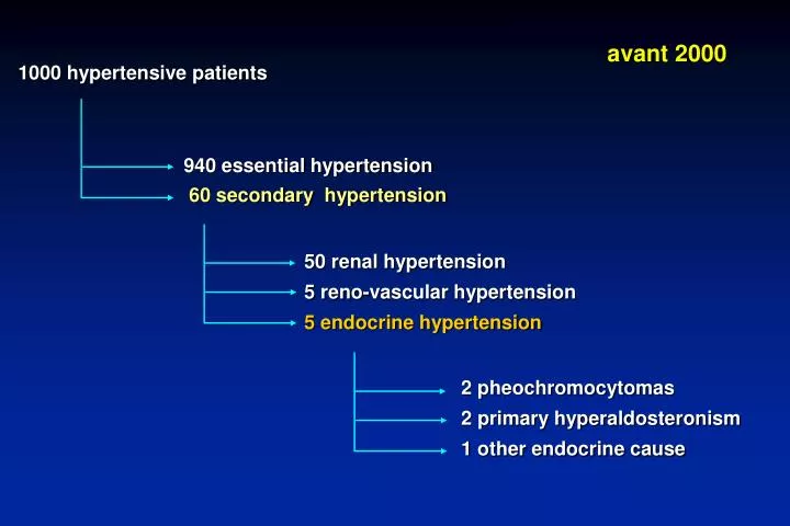
1000 hypertensive patients < 900 essential hypertension > 100 secondary hypertension 50 renal hypertension 5 reno-vascular hypertension 60 endocrine hypertension 3 pheochromocytomas 50 primary hyperaldosteronism 7 other endocrine causes après 2000
L’ hyperaldostéronisme primaire “classique” de 1965 à 2000 : le syndrome de Conn • HTA souvent sévère, résistante à un traitement classique comportant au moins 2 anti-hypertenseurs • hypokaliémie spontanée (ou facilement inductible par diurétiques ou régime riche en sel) + kaliurie > 30 mEq/24 h • hyperaldostéronisme non suppressible par une surcharge sodée ou l’administration d’un minéralocorticoïde • suppression de l’axe rénine-angiotensine (ARP ou R )
L’hyperaldostéronisme primaire (AP) revisité depuis l’an 2000 : une prévalence accrue… • plusieurs publications récentes montrent une prévalence de 5 à 20% d’AP dans une population de patients hypertendus (dépistage par le rapport aldostérone / rénine plasmatique) Lim et al, Lancet, 1999, 353: 40 Loh et al, JCEM, 2000, 85: 2854 Fardella et al, JCEM, 2000, 85: 1863… • l’hypokaliémie n’est présente que dans moins de 50% des cas ! dépistage justifié si HTA (sévère) avec normokaliémie • imagerie surrénalienne souvent négative !
Diagnostic de l’hyperaldostéronisme primaire • dépistage : • rapport aldostérone (ng/dl) (N : 5 - 15) activité rénine plasmatique (ng/ml/h) (N: 0,2 – 5) Si > 30 + aldo > 10 ng/dl (0,30 nmol/l) : forte suspicion Si > 50 + aldo > 15 ng/dl (0,40 nmol/l) : diagnostique Ce rapport varie peu avec la position, le moment du prélèvement, l’ingestion de sel, … Et les médicaments anti-hypertenseurs ??
aldostérone C. Seifarth et al.Clin. Endocrinol.,2002, 57 : 457
rénine C. Seifarth et al.Clin. Endocrinol.,2002, 57 : 457
Rapport aldostérone/rénine C. Seifarth et al.,Clin. Endocrinol.,2002, 57 : 457
Diagnostic de l’hyperaldostéronisme primaire • rapport aldostérone (ng/dl) activité rénine plasmatique (ng/ml/h) Valide si : - éventuelle hypokaliémie corrigée - apport en NaCl normal - diurétiques, -bloquants, AINS arrêtés depuis 4 semaines Faux + (rapport )Faux – (rapport ) -bloquants, clonidine diurétiques, spironolactone, IEC et Sartans AINS hypokaliémie Insuffisance rénale HTA réno-vasculaire ou maligne Patients âgés grossesse déplétion en NaCl
Diagnostic de l’hyperaldostéronisme primaire • confirmation : • rapport aldo / ARP très élevé > 50 + hyperaldostéronisme > 15 ng/dl • tests de surcharge saline • régime riche en NaCl – 3 jours (+ apports en KCl) (Mayo clinic) aldostéronurie J 3 > 14 µg/24h avec Na urinaire > 200 mEq/24h • sérum physiologique 2 litres / 4 heures iv aldostérone plasmatique > 10 ng/dl ou 0,25 nmol/l • tests de suppression • captopril (50 mg per os) • fludrocortisone (0,1 mg/6h – 4 jours) aldo > 6 ng/dl ou 0,15 nmol/l
Causes d’hyperaldostéronisme primaire • “APA” adénome surrénalien unilatéral 35% () R/ chirurgical • “BAH” hyperplasie (nodulaire) bilatérale 64% () R/ médical • Formes familiales 1% • type I – “GRA” – hyperaldostéronisme suppressible par les glucocorticoïdes – diagnostic génétique (UCL) R/ glucocorticoïdes • type II – gène non encore identifié
Mr G.DM. : CT Scan abdominal surrénale gauche Adénome surrénalien droit 9 x 1,3 cm
Hyperaldostéronisme primaire IRM abdominale montrant une hyperplasie surrénalienne bilatérale
Monsieur G. DM. : cathétérisme des veines surrénaliennes (rapports cortisol /aldo) Gradient ipsilatéral 1.9 / 0.5 = 3.8 (> 2) Gradient controlatéral 0.03 / 0.5 = 0.06 (< 1) 0.5 1.9 V.C.I. suprarénale v. surr. droite 0.03 0.3 v. rénale polaire D v. surr. gauche 0.5 0.5 0.5 0.5 v. rénale G v. rénale D V.C.I. infrarénale 0.5 Gradient de latéralisation 1.9 / 0.03 = 63 ( > 4)
Hyperaldostéronisme primaire Si jeune âge, antéc. familiaux + : exclure GRA Test de posture + Imagerie (CT / IRM) ? Dans tous les autres cas • aldostérone debout + adénome unilatéral chez patient < 40 ans Cathétérisme des veines surrénaliennes Pas de gradient G-ipsi > 2 G-contro < 1 G-latér > 5 APA BHA chirurgie R/ médical
M. Stowasser et R.D. Gordon. Primary aldosteronism Best Practice & Research Clin Endocrinology and Metabolism 2003; 591-605.
Main effects of catecholamines on the cardio-vascular system Dopamine a 1- receptors (vascular bed) + Norepinephrine Sympathetic nervous system potent vasoconstriction (300 pg/ml) Adrenal medulla tachycardia inotropic effect Epinephrine (30 pg/ml) b 1- receptors (heart) Dopamine vasodilation b 2- receptors (vascular bed) DA-receptors (vascular bed) vasodilation
Pheochromocytoma a rare tumor ... • considered as a rare cause of hypertension (0.1 - 0.2 %) • only 1 pheochromocytoma discovered in 100 investigations for paroxystic HTA
Pheochromocytoma … but underdiagnosed ! • 4-8% of incidentally-discovered adrenal masses • 50% of the cases are normotensive between spells • 75% of pheos discovered at autopsy remain undiagnosed before death (Mayo Clinic Series)
Pheochromocytoma : the rule of 10% 10% of pheochromocytomas ... • are incidentally discovered • are extra-adrenal (« paragangliomas ») • are multiple / bilateral • are or will become malignant • occur in children • are familial (probably more …) • will recur
spells of any of the following symptoms : -headaches70% - abdominal pain 25% -perspirations65% - chest pain 20% -palpitations65% - weakness 20% - pallor 45% - dyspnea 20% - nausea 35% - weight loss 15% - tremor 30% - visual disturbances 15% - anxiety 30% - polyuria, polydipsia 10% « pheo’s triad » Pheochromocytoma : clinical symptoms • paroxysmal (50%) or persistent (50%) hypertension - often severe and refractory to treatment
Pheochromocytoma : clinical symptoms • spontaneous spells or spells provoked by exercise, twisting, turning, straining, micturition, coitus, surgical procedure, delivery, abortion, …. • frequency of spells : 1 every 2-3 months 25 every day ! • duration of spells : usually 1 to 30 minutes
Pheochromocytoma : who should be screened for? • paroxysmal or markedly fluctuating hypertension • “spells” of any symptoms + hypertension • pheo’s triad [headaches + sweating + palpitations] • refractoriness to conventional anti-hypertensive treatment • (orthostatic) hypotension alternating with hypertension • paradoxical hypertensive response to beta-blockers • adrenal incidentaloma • personal history of a predisposing disease (von-Hippel-Lindau, MEN2, neurofibromatosis, …) • familial history of pheochromocytoma, MEN2 syndrome, ...
Pheochromocytoma : how to screen ? Biochemical test reference value sensitivity specificity urinary NMN + MN > 1.2 mg/24h 90-95% 98% urinary NE + E > 200 µg/24 h 80-85% 98% urinary VMA > 11 mg/24h 30-35% 99-100% urinary NE + E > 200 µg/24h or urinary NMN + MN > 1.2 mg/24h plasma NE + E > 1000 pg/ml 88-90% 90% > 2000 pg/ml low 100% 98% 98% In most cases, urinary mets + cats are diagnostic No test has 100% sensitivity + 100% specificity !
Pheochromocytoma : value of fractionated plasma metanephrines Sensitivity = 99% specificity = 85% pheo controls pheo controls May be useful if high suspicion A. Sawka et al., JCEM, 2003
Pheochromocytoma : the clonidine suppression test Pheochromocytoma (n=10) Non-pheochromocytoma (n=15) 20000 10000 8000 6000 4000 2000 Plasma norepinephrine (pg/ml) 1000 800 600 400 200 100 Basal Post-clonidine
Pheochromocytoma : how to localize ? Sensitivity Specificity CT-Scan 97% 70% MRI 99% 80% 131I-MIBG 80% 100% 111In-octreoscan 70% 95% PET-Scan ? ? (FDG, fluorodopamine)
Pheochromocytoma : magnetic resonance imaging T1-weighed image T2-weighed image
Pheochromocytoma : 131-I MIBG Scanning Posterior Lateral R Ant R
Familial pheochromocytoma • multiple endocrine neoplasia type II (MEN IIa, MEN IIb) • von Hippel-Lindau disease (VHL type 2) • von Recklinghausen’s neurofibromatosis • hereditary paraganglioma • familial isolated pheochromocytoma
- - - - - Clinical suspicion of pheochromocytoma high low 2-fold increase U-cats U-mets > 1.2 mg/24 h positive clonidine test ( plasma mets) 24h-urinary mets+cats - 24h-urinary mets+cats - clonidine test - (plasma mets) + + Other causes of spells? Abdominal MRI / CT 131I-MIBG recheck after a spell do a provocative test ? + + negative Pheochro- mocytoma + Other causes of spells? Whole body MRI octreo-scan PET-scan Other causes of hypercate- cholaminemia Preparation + surgery
Maiter D. Pheochromocytoma: a paradigm for catecholamine-mediated hypertension Acta Clin Belg, 2004, 59: 209-219.