Download
1 / 25
340 likes | 1.36k Views
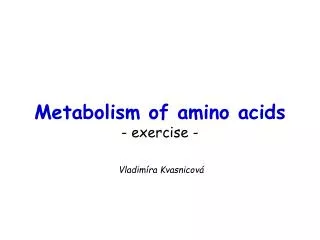
Pavla Balínová. Metabolism of amino acids, purine and pyrimidine bases. Amino acids (AAs). Sources of AAs: diet synthesis de novo protein degradation. dietary proteins proteosynthesis body proteins AAs pool N-compound synthes.

E N D
Pavla Balínová Metabolism of amino acids, purine and pyrimidine bases
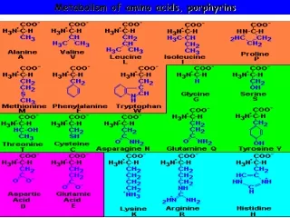
Amino acids (AAs) Sources of AAs: • diet • synthesis de novo • protein degradation dietary proteins proteosynthesis body proteins AAs pool N-compound synthes. de novo biosynthesis degradation (E,glc,fat)
Essential AA Nonessential AA Arg Ala His Asn Ile Asp Leu Cys Lys Gln Met Glu Phe Gly Thr Pro Trp Ser Val Tyr Biosynthesis of amino acids (AA) Humans can synthesize only 10 of the 20 AA. Essential AA = AA that cannot be synthesized „de novo“. They must be obtained from diet. Nonessential AA: Ala is synthesized from pyruvate. Cys is synthesized from Met and Ser. Tyr is formed by hydroxylation from Phe.
Synthesis of AAs in a human body- 5 substrates - • oxaloacetate→ Asp, Asn • -ketoglutarate→ Glu, Gln, Pro, (Arg) • pyruvate → Ala • 3-phosphoglycerate→ Ser, Cys, Gly • Phe→ Tyr
Synthesis of thyrosine from phenylalanine Figure is found at http://themedicalbiochemistrypage.org/amino-acid-metabolism.html#tyrosine
Formation of activated methionine= S-adenosylmethionine (SAM) SAM is used as –CH3 group donorin metabolic methylations Figure is found at http://themedicalbiochemistrypage.org/amino-acid-metabolism.html#cysteine
Synthesis ofCysfrom Met and Ser Figure is found at http://themedicalbiochemistrypage.org/amino-acid-metabolism.html#cysteine
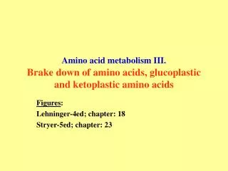
Degradation of AA 20 different multienzyme sequences exist for catabolism of AAs. All common 20 AAs are converted to only 7 compounds: • pyruvate • acetyl-CoA • acetoacetyl-CoA • α-ketoglutarate • succinyl-CoA • fumarate • oxaloacetate
Three types of reactions are typical for degradation of AAs: • Transamination 2. Deamination 3. Decarboxylation
Transamination = an exchange of –NH2 between amino acid and α-ketoacid These reactions are catalyzed by transaminases (aminotransferases). Most of them require α-ketoglutarate as an acceptor of –NH2. Coenzyme of transaminases: pyridoxal phosphate (vit. B6 derivative) Figure is found at http://web.indstate.edu/thcme/mwking/nitrogen-metabolism.html
Aminotransferases (transaminases)important in medicine Alanine aminotransferase (ALT) Aspartate aminotransferase (AST) Figure was adopted from Devlin, T. M. (editor): Textbook of Biochemistry with Clinical Correlations, 4th ed. Wiley‑Liss, Inc., New York, 1997. ISBN 0‑471‑15451‑2
Deamination e. g.oxidative deamination of Glu Glu→ α-ketoglutarate by glutamate dehydrogenase Figure is found at http://www.sbuniv.edu/~ggray/CHE3364/b1c25out.html
Decarboxylation → primary amines a)decarboxylation of His → histamine b) decarboxylation of Trp → serotonin c) decarboxylation of Tyr → epinephrine and norepinephrine d) decarboxylation of Glu → GABA (γ-aminobutyrate) Figure is found athttp://www.sbuniv.edu/~ggray/CHE3364/b1c25out.html
Ammonia transport and detoxification Glutamine (Gln) is the major transport form of ammonia. Figure is found at http://www.sbuniv.edu/~ggray/CHE3364/b1c25out.html
Glucose-Alanine cycle Figure is found at http://www.sbuniv.edu/~ggray/CHE3364/b1c25out.html
Urea cycle (ornithine cycle) • substrates: NH4+, HCO3-, Asp, ATP • product: urea • function: synthesis of non-toxic urea • subcellular location: mitochondria and cytosol • organ location: liver • regulatory enzyme: carbamoyl phosphate synthetase I
Urea cycle (ornithine cycle) l Figure is found at http://web.indstate.edu/thcme/mwking/nitrogen-metabolism.html
The fate of carbon skeletons of AA during catabolism • The strategy of the cell is to convert carbon skeletons to compounds useful in gluconeogenesis or CAC. • Glucogenic AAs = AA that can form any of intermediates of carbohydrate metabolism Gly, Ala, Ser, Cys, Thr → pyruvate Glu, Pro, Arg, His → Glu → α-ketoglutarate Met, Ile, Val → succinyl-CoA • Ketogenic AAsare converted to acetyl-CoA and acetoacetyl-CoA. They yield ketone bodies. Leu, Lys ● Glucogenic-ketogenic AAs = Thr, Phe, Tyr, Ile
Figure is found at http://www.biocarta.com/pathfiles/glucogenicPathway.asp Figure is found
De novo synthesis of purine nucleotides Figure is found at http://web.indstate.edu/thcme/mwking/nucleotide-metabolism.html
Important notes about biosynthesis of purine nucleotides • Subcellular location: cytoplasm • PRPP = phosphoribosyl pyrophosphate is derived from ribose-5-P • IMP = inosine monophosphate serves as the common precursor of AMP and GMP synthesis • Gln, Gly, Asp are donors of C and N atoms • CO2 is a source of C • C1 units are transferred via tetrahydrofolate „Salvage pathway“: • purines from normal turnover of cellular NA can be converted to nucleoside triphosphates • substrates: purine bases, PRPP, ATP
Degradation of purine nucleotides → uric acid is formed by enzyme xanthine oxidase
De novo synthesis of pyrimidine nucleotides Figure is found at http://web.indstate.edu/thcme/mwking/nucleotide-metabolism.html
Important notes about synthesis of pyrimidine nucleotides • Carbamoyl phosphate is formed from Gln and CO2 (2 ATP are consumed). This reaction is catalyzed by carbamoyl-P synthetase II (cytosolic enzyme) = regulatory step • pathway occurs in cytoplasm but formation of orotate occurs in mitochondrion→ orotate is linked by PRPP → OMP → UMP • UMP→ UTP →CTP TTP ● UTP inhibits regulatory enzyme, activator is PRPP „Salvage pathway“: ● pyrimidine nucleosides are phosphorylated (ATP) to nucleotides TTP
Degradation of pyrimidine bases → β-amino acids are formed