Download

1 / 143
1.53k likes | 2.02k Views
Head Trauma. D r : Zohair AlAseri FRCPc, Emergency Medicine FRCPc, Critical Care Medicine FCEM UK Chairman, Department of Emergency Medicine King khalid University Hospital, Riyadh, KSA. Head Trauma. 35 year old male involved in motor vehicle collision Presented with GCS of 8

E N D
Head Trauma Dr: Zohair AlAseri FRCPc, Emergency Medicine FRCPc, Critical CareMedicine FCEM UK Chairman, Department of Emergency Medicine King khalid University Hospital, Riyadh, KSA.
Head Trauma 35 year old male involved in motor vehicle collision Presented with GCS of 8 And BP of 85/32, HR of 140 What is your 1st line of treatment 1—intubate 2---IV fluid 3---CT scan to exclude intracranial bleed 4---hypervetilation
35 year old male involved in motor vehicle collision Presented with GCS of 8, smell of ethanol And BP of 85/32, HR of 140 What is most likely cause of his hypotension 1---sever head trauma 2---hypovolemia 3---intoxication
Head Trauma • Minor head trauma (Glasgow Coma Scale [GCS] score of 14 to 15) or presence of any intracranial contusion, hematoma, or laceration • Moderate head injuries (GCS of 9 to 13) • Severe head injuries (GCS of 8 or less)
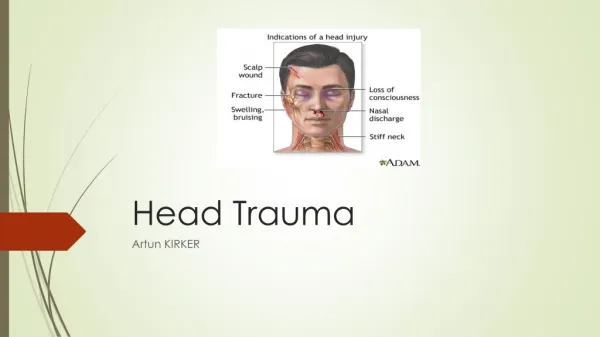
Head Trauma • External physical signs • not always present in the patient who has sustained serious underlying traumatic brain injury (TBI).
Head Trauma Cerebral Hemodynamics Blood-Brain Barrier. • The normal pressure exerted by the CSF is 65 to 195 mm H2O or 5 to 15 mm Hg.
Head Trauma Cerebral Hemodynamics Blood-Brain Barrier. • The blood-brain barrier (BBB) maintains the microenvironment of the brain tissue. • Extracellular ion and neurotransmitter concentrations are regulated by movement across BBB. • The brain has an extremely high metabolic rate, using approximately 20% of the entire oxygen volume consumed by the body So it requires about 15% of the total cardiac output.
Head Trauma Cerebral Hemodynamics Blood-Brain Barrier. • The brain has an extremely high metabolic rate • using approximately 20% of the entire oxygen volume consumed by the body • it requires about 15% of the total cardiac output.
Head Trauma Cerebral Hemodynamics Blood-Brain Barrier. • Hypertension, alkalosis, and hypocarbia promote cerebral vasoconstriction • hypotension, acidosis, and hypercarbia cause cerebral vasodilation.
Head Trauma Cerebral Hemodynamics Pco2 Over time, injured vessels lose their responsiveness to hypocarbia become vasodilated. increased brain swelling and mass effect.
Head Trauma Cerebral Hemodynamics Po2 Low Po2 ----- cerebral vessels dilate vasogenic edema. So hypoxia should be treated
Head Trauma Cerebral Perfusion Pressure & BP CPP is estimated as MAP minus ICP. • CBF remains constant when CPP is 50 to 160 mm Hg. • If CPP falls below 40 mm Hg, the autoregulation of CBF is lost----ischemia So hypotension & increased ICP should be controlled
Head Trauma Primary and Secondary Brain Injury • Primary ---- damage that occurs at the time of head trauma. • it causes permanent mechanical cellular disruption and microvascular injury.
Head Trauma Secondary Brain Injury • Secondary brain injury results from intracellular and extracellular derangements All currently used acute therapies for TBI are directed at reversing or preventing secondary injury.
Head Trauma Secondary Brain Injury Influence the outcome Common secondary systemic insults in trauma patients include • Hypotension • Hypoxia • Anemia. • hypercarbia, hyperthermia, coagulopathy, and seizures.
Head Trauma All Bad Secondary Brain Injury • Hypotension doubles the mortality • Hypoxia, defined as a Po2 less than 60 mm Hg • AnemiaWhen anemia (hematocrit less than 30%) occurs in patients with severe head injury, the mortality rate increases Brain Trauma Foundation, American Association of Neurological Surgeons, Joint Section on Neurotrauma and Critical Care : Guidelines for the management of severe traumatic brain injury. J Neurotrauma 2000; 17:471.
Contributing events in the pathophysiology of secondary brain injury.
Head Trauma Altered Levels of Consciousness Hallmark of brain insult Causes • hypoxic • Hypotension • intoxication consumed before the injury.
35 year old male involved in motor vehicle collision Presented with GCS of 8, smell of ethanol And BP of 170/32, HR of 40 and bouts of irregular breathing Your next action will be 1—consult NS 2—admit for evaluation 3—manitol 4---atropine
Head Trauma Cushing's Reflex • Progressive hypertension associated with bradycardia and diminished respiratory effort • D/T acute, potentially lethal rises in ICP.
Head Trauma Cushing's Reflex Triad How frequent is seen in cushing reflex • The full triad of hypertension, bradycardia, and respiratory irregularity is seen in only one third of cases of life-threatening increased ICP.
Head Trauma Cerebral Herniation • When increasing ICP cannot be controlled, the intracranial contents shift and herniate through the cranial foramen. • Herniation can occur within minutes to days • mortality approaches 100% without rapid implementation of temporizing emergency measures and definitive neurosurgical therapy.
Head Trauma Uncal Cerebral Herniation • The most common • a form of transtentorial herniation. • hematomas in the lateral middle fossa or the temporal lobe.
Head Trauma Uncal Cerebral Herniation • Third cranial nerve is compressed; ipsilateral anisocoria, ptosis, impaired extraocular movements, and a sluggish pupillary light reflex • As the herniation progresses, compression of the ipsilateral oculomotor nerve eventually causes ipsilateral pupillary dilation and nonreactivity.
Head Trauma Uncal Cerebral Herniation • Contralateral Babinski's • Contralateral hemiparesis • Decerebrate posturing eventually occurs; • LOS & change in respiratory pattern, and cv system. • Herniation that is uncontrolled progresses rapidly to brainstem failure, cardiovascular collapse, and death. Kernohan's notch syndrome When hemiparesis is detected ipsilateral to the dilated pupil and the mass lesion, it causes false-localizing motor findings
Head Trauma CLINICAL FEATURES, History • mechanism • comorbid factors. • Past medical history, • Medications • level of consciousness, course • Witnessed posttraumatic seizures • apnea
Head Trauma Acute Neurologic General Examination • Identification of life-threatening injuries and of neurologic changes in the immediate posttrauma period. • mental status • GCS • pupillary size • Responsiveness • motor strength and symmetry. • neurologic assessment in the immediate posttrauma period serves as a baseline
Head Trauma Glasgow Coma Scale • The GCS assesses a patient's best eye, verbal, and motor responsiveness. • limitations. • Hypoxia, hypotension, and intoxication can falsely lower the initial GCS. • Intubation • Periorbital edema • Extremity fractures • Decisions on continued resuscitation should not be based on the initial GCS
Head Trauma Pupillary Examination • must be done early • A large fixed pupil suggests herniation syndrome Limitations: Traumatic mydriasis, resulting from direct injury to the eye and periorbital struc-tures, may confuse the assessment of the pupillary responsiveness. Atropine///???
Head Trauma Motor Examination: Posturing • A false-localizing motor examination • Kernohan's notch syndrome • occult extremity trauma • spinal cord injury • nerve root injury • motor movement should be elicited by application of noxious stimuli.
Head Trauma Motor Examination: Posturing • Decorticate posturing • implies injury above the midbrain. • Decerebrate posturing • is the result of a more caudal injury and therefore is associated with a worse prognosis.
Head Trauma Brainstem Function • Respiratory pattern • Pupillary size • Eye movements • The oculocephalic response • The oculovestibular response (cold water calorics) • (CN) examination is often limited to the pupillary responses (CN III), • Gag reflex (CNs IX and X) • Corneal reflex (CNs V and VII). • Facial symmetry (CN VII) with noxious stimuli.
Clinical Characteristics of Basilar Skull Fractures Cranial nerve deficits
Head Trauma Clinical prognostic indicators • initial motor activity • pupillary responsiveness • Age • premorbid condition • secondary systemic insult • The prognosis cannot be reliably predicted by the initial GCS or initial CT scan.
Head Trauma MANAGEMENT, Laboratory Tests • complete blood count • Electrolytes • Glucose • coagulation studies. • ECG
Head Trauma MANAGEMENT, Neuroimaging • non–contrast-enhanced head CT scan. • Emergency management decisions are strongly influenced by these acute CT scan findings. MRI is better than CT in detecting • posttraumatic ischemic infarctions • subacute nonhemorrhagic lesions • contusions • axonal shear injury • lesions in the brainstem or posterior fossa
Head Trauma MANAGEMENT Out-of-Hospital Care • The goals of the out-of-hospital management are necessary airway interventions to prevent hypoxia • establishing intravenous (IV) access to treat trauma-related hypotension. • GCS • pupillary responsiveness and size • level of consciousness • motor strength and symmetry.
Head Trauma MANAGEMENT • All head-injured patients should have a cardiac monitor as they are transported from the accident scene.
Head Trauma MANAGEMENT, Airway • Rapid sequence intubation (RSI) • a brief neurologic examination before RSI • Lidocaine (1.5 to 2 mg/kg IV push) may help as premedication
Head Trauma MANAGEMENT, Airway • Thiopental may also be effective but should not be used in hypotensive patients. • Etomidate (0.3 mg/kg IV) • a short-acting sedative-hypnotic agent • beneficial effects on ICP by reducing CBF and metabolism. • minimal adverse effects on blood pressure
Head Trauma MANAGEMENT, Airway • Combinations of ketamine-midazolam or ketamine-sufentanil have recently been shown to be comparable in maintaining ICP and CPP in patients with severe head injury receiving mechanical ventilation Bourgoin A , Albanese J , Wereszczynski N , et al: Safety of sedation with ketamine in severe head injury patients : Comparison with sufentanil . Crit Care Med 2003 ; 31 : 711–717
Head Trauma MANAGEMENT, Airway • Propofol : the preferred sedating agent based on its short duration of action, facilitating serial neurologic evaluations. • Propofol-induced hypotension may occur, and it should be titrated carefully McKeage K , Perry CM : Propofol : A review of its use in intensive care sedation of adults . CNS Drugs 2003 ; 17 : 235–272
Head Trauma MANAGEMENT, Hypotension • rarely caused by head injury in adult • spinal cord injury, neurogenic hypotension may occur. • fluids do not produce clinically significant increases in ICP; SO should never be withheld in the head trauma patient with hypovolemic hypotension for fear of increasing cerebral edema and ICP • normal saline or lactated Ringer's solution or hypertonic saline
Head Trauma MANAGEMENT, Hypotension • Ract C , Vigue B : Comparison of the cerebral effects of dopamine and norepinephrine in severely head-injured patients . Intensive Care Med 2001 ; 27 : 101–106 • Steiner LA , Johnston AJ , Czosnyka M , et al: Direct comparison of cerebrovascular effects of norepinephrine and dopamine in head-injured patients . Crit Care Med 2004 ; 32 : 1049–1054 • Johnston AJ , Steiner LA , Chatfield DA , et al: Effect of cerebral perfusion pressure augmentation with dopamine and norepinephrine on global and focal brain oxygenation after traumatic brain injury . Intensive Care Med 2004 ; 30 : 791–797 Norepinephrine Vasopressor of choice if fluid is not doing the job
Head Trauma MANAGEMENT, analgesia • There is no real preference for one analgesic agent over another • The key factor is that arterial hypotension secondary to excessive doses of a sedative/analgesic should be avoided and is more likely to occur in patients with underlying hypovolemia.
Head Trauma MANAGEMENT, Hyperventilation • only in patients demonstrating neurologic deterioration. • onset of action is within 30 seconds • peaks within 8 minutes after the Pco2 drops to the desired range. • Pco2 should not fall below 25 mm Hg
Head Trauma MANAGEMENT, Mannitol • For increased ICP • Mannitol (0.25 to 1 g/kg) • works within minutes • peak about 60 minutes after bolus administration. • The ICP-lowering effects of a single bolus may last for 6 to 8 hours.
Head Trauma MANAGEMENT, Mannitol • It is an effective volume expander • It also promotes CBF by reducing blood viscosity and microcirculatory resistance. • It is an effective free radical scavenger, Limitation • renal failure or hypotension if given in large doses. • paradoxical effect of increased bleeding into a traumatic lesion by decompressing the tamponade effect of a hematoma.