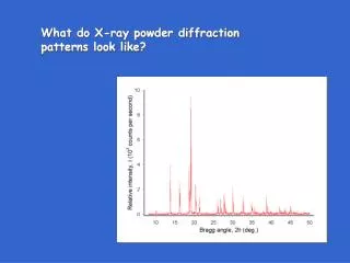
What do X-ray powder diffraction patterns look like?
What do X-ray powder diffraction patterns look like?. Powder patterns - what information available in pattern?. 1. peak positions peak intensities - get crystal structure peak shape background structure. Intensities give atom positions. Intensities.

What do X-ray powder diffraction patterns look like?
E N D
Presentation Transcript
What do X-ray powder diffraction patterns look like?
Powder patterns - what information available in pattern? 1. peak positions peak intensities - get crystal structure peak shape background structure Intensities give atom positions
Intensities X-rays scattered by electrons - electrons are in atoms Scattering power f of atom at = 0° is no. electrons (atomic no.) x scattering power of one e—
Intensities Because of path length difference, scattering power decreases as increases Decrease is gradual since path length difference is small compared to Atomic scattering factor f plotted as a function of (sin /. f values given in various tables, and as analytical functions
Intensities Now think of atoms in unit cells of a lattice Waves reflected (diffracted) from the same atoms at the same x, y, z positions in all unit cells will be in phase Thus, need to consider scattering in only one unit cell Out-of-phaseness depends on: a. relative positions of atoms xj, yj, zj b. diffraction angle As before, the scattered waves out of phase But distances between the atoms much larger than between e— s
The scattering power for all atoms in unit cell obtained by adding up all scattered waves. This is the structure factor or structure amplitude Intensities Wave from each atom has: amplitude - ƒ phase factor - exp (2πij) = exp (2πi(hxj + kyj + lzj) xj, yj, zj are positions h, k, l related to diffraction angle
Intensities Since Fhkl is an amplitude Ihkl ~ |Fhkl|2 In general, F is imaginary ........ so F = A + iB and F*F = (A - iB) (A + iB)
Intensities Simple example calculation: Cu: Fm3m a = 3.614 Å Cu atoms in 4a - (000) (1/2 0 1/2) (1/2 1/2 0) (0 1/2 1/2) Equipoint 4a —> 4 atoms/cell —> 4 terms: Fhkl = ƒCu (e0 + eπi(h + l) + eπi(h + k) + eπi(k + l)) Since ei = cos + i sin (Euler's rule) Fhkl = 0 or 4ƒCu
Then Fhkl = ∑ ƒj {(exp (2πi(hxj + kyj + lzj) + exp (2πi(h(xj + 1/2) + k(yj + 1/2) + l(zj + 1/2)))} N/2 j = 1 Since eA + B = eA eB Fhkl = ∑ƒj (exp (2πi(hxj + kyj + lzj)))(1 + eπi(h + k + l)) N/2 j = 1 Intensities Derivation of extinction rule for I centering: For every atom at (xj, yj, zj), there must be an atom at (xj + 1/2, yj + 1/2, zj + 1/2)
Since eA + B = eA eB Fhkl = ∑ƒj (exp (2πi(hxj + kyj + lzj)))(1 + eπi(h + k + l)) N/2 j = 1 Intensities Derivation of extinction rule for I centering Every term in sum contains 1 + eπi(h + k + l) = 1 + cos (π(h + k + l)) + i sin (π(h + k + l)) —> 0 when h + k + l = odd number Extinction rule for I centering: (hkl), h + k + l = 2n
Intensities A slightly more complex structure: HoZn2 is I 2/m 2/m 2/a, with a = 4.456 ± 1, b = 7.039 ± 3, c = 7.641 ± 5Å Ho in 4e (0,1/4,z) (0,3/4,z) + I , z = 0.5281 ± 4 Zn in 8h (0,y,z) (0,y,z) (0,1/2 + y,z) (0,1/2-y,z) + I, y = 0.0410 ± 9, z = 0.1663 ± 8 F(hkl) = fHo (exp (2πi(k/4 + l(0.5281))) + exp (2πi(3/4k + l(0.4719))) + exp (2πi(1/2h + 3/4k + l(0.0281))) + 1 more term) + fZn (exp (2πi(k(0.0410) + l(0.1663))) + 7 more terms)
Example - cubic {100} = (100), (010), (001), (100), (010), (001) p = 6 {110} = (110), (101), (011),, (110), (101), (011), (110), (101), (011), (110), (101), (011) p = 12 Intensities Now Ihkl = scale factor • p • LP • A • |Fhkl|2 • e–2M p = multiplicity accounts for differing probabilities that symmetry equivalent planes (hkl) will reflect
Intensities Now Ihkl = scale factor • p • LP • A • |Fhkl|2 • e–2M LP = Lorentz-polarization factor P accounts for polarization state of incident beam (in most powder x-ray diffractometers, unpolarized) Lorentz factor corrects for geometrical broadening of reflections as 2 increased
Which reflection is more intense? Intensities Now Ihkl = scale factor • p • LP • A • |Fhkl|2 • e–2M Lorentz factor corrects for geometrical broadening of reflections as 2 increased
Intensities Now Ihkl = scale factor • p • LP • A • |Fhkl|2 • e–2M LP = Lorentz-polarization factor LP = (1 + cos2 2q)/sin2q cos q
Intensities Now Ihkl = scale factor • p • LP • A • |Fhkl|2 • e–2M A = absorption factor In standard X-ray powder diffractometers, when specimen is dense and thick, A is considered constant over all 2
I(high T) e–2M(high T) I(low T) e–2M(low T) 1 e2M(high T) - 2M(low T) = = Intensities Now Ihkl = scale factor • p • LP • A • |Fhkl|2 • e–2M(T) e–2M(T) = temperature factor (also called Debye-Waller factor) 2M(T) = 16π2 m(T)2(sinq)2/l2 m2= mean square amplitude of thermal vibration of atoms direction normal to planes (hkl)
Measure reflection intensities, get F-values, calculate electron density distribution from Intensities —> crystal structure So, OK, how do we do it? Outline of procedure: Measure reflection positions in x-ray diffraction pattern - index, get unit cell type and size, possible space groups Measure density, if possible, to get number formula units/unit cell (N) density = N x formula wt/cell volume x Avogadro's no.
anthracene Intensities —> crystal structure Electron density distribution tells where the atoms are (XYZ) is plotted and contoured to show regions of high electron density
√Ihkl/K = √(Ahkl2 + Bhkl2) So, can't use Ihkls directly to calculate Fhkls and (XYZ)!! Intensities —> crystal structure But WAIT!!! Ihkl = K |Fhkl|2 = K Fhkl* x Fhkl = K (Ahkl - iBhkl) (Ahkl + iBhkl) = K (Ahkl2 + Bhkl2) Many techniques for using Ihkls to determine atom positions have been developed, most of which, at some stage, involve formulating a model for the crystal structure, and then adjusting it to fit the intensity data
Powder patterns - what information available in pattern? 1. peak positions peak intensities peak shape - peak broadening background structure Two effects
X-rays at non-angles won't reflect; every reflected ray has a mate deeper in the crystal which is 180° out of phase with it - reflection is narrow Peak broadening Here, have set of planes reflecting in-phase at (Bragg condition) Small crystallite size broadens reflections - becomes significant below 1 micron
Peak broadening Microstrain & chemical inhomogeneity distort the structure so that interplanar distances not constant - vary a little from an average value - broadens peaks
Must subtract broadening due to instrument. Measure peak width from suitable standard (ex: LaB6 - NIST SRM 660) Peak broadening If broadening due to small crystallite size only, simple technique to determine that size, L, from the breadth, B Scherrer eqn.: Bsize = (180/π) (K/ L cos ) (K - 0.9, usually) Btot2 = Binstr2 + Bsize2
Peak broadening If broadening due to small crystallite size only, simple techique to determine that size, L, from the breadth, B Scherrer eqn.: Bsize = (180/π) (K/ L cos ) (K - 0.9, usually) Btot2 = Binstr2 + Bsize2 104Å Bsize = (180/π) (1.54/ 104 cos 45°) = 0.0125° 2 103Å Bsize = 0.125° 2 102Å Bsize = 1.25° 2 10Å Bsize = 12.5° 2
Peak broadening If broadening due to small crystallite size & microstrain, simple technique to determine size, L, & strain, <>, from the breadth, B Williamson-Hall method: strain broadening - Bstrain = <>(4 tan ) size broadening - Bsize = (K/ L cos ) (Bobs − Binst) = Bsize + Bstrain (Bobs − Binst) = (K / L cos ) + 4 <ε>(tan θ) (Bobs − Binst) cos = (K / L) + 4 <ε>(sin θ)
Peak broadening If broadening due to small crystallite size & microstrain, simple technique to determine size, L, & strain, <>, from the breadth, B Williamson-Hall method: (Bobs − Binst) cos = (K / L) + 4 <ε>(sin θ)
Peak broadening If broadening due to small crystallite size & microstrain, can use other more complex methods for analysis: Warren-Averbach double Voigt x-ray tracing
Obvious structural information in back-ground - due to non-crystalline character of material (disorder) Powder patterns - what information available in pattern? 1. peak positions peak intensities - get crystal structure peak shape background structure