Download

1 / 70
700 likes | 717 Views
Introduction to Cardiovascular System, What is Blood? Functions of Blood, pH , Composition of Blood, hemostasis , Blood coagulation, Hemopoieses and S.P gravity. Dr. Tariq Hussain Assistant Professor Pharmacology and Toxicology, College of Veterinary and Animal Sciences, Jhang.

E N D
Introduction to Cardiovascular System, What is Blood? Functions of Blood, pH , Composition of Blood, hemostasis, Blood coagulation, Hemopoieses and S.P gravity. Dr. Tariq Hussain Assistant Professor Pharmacology and Toxicology, College of Veterinary and Animal Sciences, Jhang.
Introduction to Cardiovascular System • Cardiovascular System • System made up of blood vessels, blood and heart. Major function is to transport nutrients, gases and hormones to the cells and pick up wastes from cells to transport them to the excretory organs of body where they are excreted • Lymphatic System • Network of vessels that return the fluid escaped from blood vessels back to the bloodstream • Includes lymphocytes, lymphoid tissue and lymphoid organs which fight infections and give immunity to disease • Circulatory System • Together the cardiovascular system and lymphatic system make up the circulatory system
What is Blood? • Blood is a connective tissue • Its volume is 5-6 L in males and 4-5 L in females (in humans) • It is slightly alkaline in nature • The pH of blood ranges from 7.35 to 7.45 • Its color varies from bright to dark red • It has a salty metallic taste
Functions of Blood 3 major functions • Transportation • Regulation • Protection
Conti… Functions of Blood Transportation: • Transportation of dissolved Gases Red blood cells or erythrocytes transport Oxygen from lungs to cells and Carbon dioxide from cells to lungs • Transportation of Nutrients Blood absorb nutrients from digested foods in gastrointestinal tract and transport to all the cells in body
Conti… Functions of Blood • Transportation of metabolic waste Metabolic wastes, excess water and ions , and other molecules not needed by the body are carried by the blood to the kidneys and excreted in the urine
Conti… Functions of Blood Regulation: • Regulation of Hormones Blood carries hormones from their site of origin to distant target tissues , where they perform the regulatory functions • Regulation of Temperature Blood is responsible to carry body heat to the surface in high temperature environment as well as to keep body heat in within low temperature environment • Regulation of pH and Electrolytes Blood regulates the pH and electrolyte composition of the interstitial fluids.
Conti… Functions of Blood Protection: • Protection against Bleeding The clotting mechanism protects against blood loss when vessels are damaged • Protection against Infectious Diseases The immune function of blood is performed by the leukocytes that protects against many disease causing agents
Conti….. Composition of Blood Major Components: • Plasma Fluid portion containing Fibrinogen • Formed elementsBlood cells (RBCs, WBCs and Platelets)
Conti….. Composition of Blood Plasma: • Straw-colored, sticky fluid portion of blood • Approximately 90% water • Contains: • Ions – Na+ and Cl- • Nutrients – sugars, amino acids, lipids, cholesterol, vitamins and trace elements • Three main proteins - Albumin (60%), globulin (35%), fibrinogen (4%) • Dissolved Gasses – including O2 and CO2 • Waste Products – other protein wastes such as urea and bilirubin
Cont….. Composition of Blood Formed Elements of Blood: • RBCs (Erythrocytes) • WBCs(Leukocytes) • Granulocytes • Neutrophils • Eosinophils • Basophils • Agranulocytes • Lymphocytes • Monocytes • Platelets (Thrombocytes)
Cont….. Composition of Blood Erythrocytes – Red Blood Cells (RBCs): • Oxygen-transporting cells • 7.5 µm in diameter (diameter of capillary 8 – 10µm) • Most numerous of the formed elements • Females: 4.3 – 5.2 million cells/cubic millimeter • Males: 5.2 – 5.8 million cells/cubic millimeter • Made in the red bone marrow in long bones, cranial bones, ribs, sternum, and vertebrae • Average lifespan 100 – 120 days
Cont….. Composition of Blood Structure And Function of RBCs: • Have no organelles or nuclei except RBCs of poultry • Hemoglobin – oxygen carrying protein • Each RBC has about 280 millionhemoglobin molecules • Biconcave shape – 30% more surface area
Cont….. Composition of Blood Structure of Hemoglobin: • RBC’s pass through capillary beds in single file. • Hemoglobin is made of four polypeptides: Two Alpha and two Beta that contain a heme unit • Each heme can carry an O2 • There are about 280 million hemoglobin molecules in each RBC • Other molecules such as CO2 and NO are also carried by hemoglobin
Cont….. Composition of Blood Changes in hemoglobin: • Anemia: A deficiency of RBCs, which can be caused by either too rapid loss or slow production. • Blood loss Anemia: Due to hemorrhage, plasma is replaced in 1-3 days, but, RBC replacement takes longer. • Microcytic Hypochromic Anemia: Low levels of hemoglobin in RBCs due to chronic blood loss resulting in low Fe3+ levels in newly produced RBCs. • Aplastic Anemia: Decreased RBC production in bone marrow due to chemical, drug, or radiation exposure. • Pernicious Anemia: Chronic illness caused by impaired absorption of Vitamin B-12 because of a lack of intrinsic factor (IF) in gastric secretions. Vitamin B12, in turn, is necessary for the formation of red blood cells.
Cont….. Composition of Blood • Hemolytic Anemia: Different abnormalities of RBCs that make RBCs fragile and rupture easily. • Hereditary Spherocytosis: RBC develop as small spherical cells rather than being biconcave. These spherical cells easily rupture by slight compression. • Sickle-cell Anemia: Genetic mutation causing abnormal beta chains. When this hemoglobin is exposed to low O2 concentrations, it precipitates into long crystals that cause the cells to become sickle-shaped.
Cont….. Composition of Blood Abnormal Hematocrits: • Polycythemia: Increased production of RBCs is known as Polycythemia. • Physiologic Polycythemia: Increase in RBC production due to hypoxic tissues, like what occurs at high altitudes. • Polycythemia Vera: Genetic mutation in the hemocytoblastic cell line that increases RBC production. Hematocrit values can reach 70%
Cont….. Composition of Blood Leukocytes – White Blood Cells (WBCs): • Protect the body from infectious microorganisms • 4,800 – 11,000/cubic millimeter • Diapedesis – circulating leukocytes leave the capillaries and comes to the interstitial fluid. • WBCs are larger than RBCs and have a nucleus • Most produced in bone marrow • Lifespan of 12 hours to several years
Cont….. Composition of Blood Granulocytes: • Neutrophils: – Most numerous among WBCs, 40 – 70% of all WBCs. – First line of defense against bacterial infection. – Size: 10 – 12 µm in diameter. – Nucleus: has 2– 6 lobes. – Upon tissue injury, rapidly accumulate within interstitial fluid or invaded area. • Phagocytize and destroy bacteria. • Chemotactic factor is a chemical messenger for Diapedesis • Neutrophilia during bacterial infection, Neutropenia
Cont….. Composition of Blood • Eosinophils: – Compose 1 – 4% of all WBCs – Size: 10 –12 µm in diameter, 2-3 lobed nucleus – Cytoplasm filled with large red granules • Play roles to regulate histamine allergic reactions • By removing antigen antibody complex which stimulate allergic reaction. • By inhibiting synthesis of chemical mediators e.g histamine • Destroy some parasitic infections
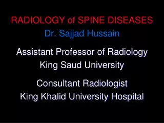
Cont….. Composition of Blood • Basophils: – They are least numerous blood cells, about 0 – 1% of all leukocytes. – 8- 10 µm in diameter, usually two lobed nucleus – Cytoplasm filled with large blue purple granules – During allergic reactions the granules liberate Heparin: Prevents blood clotting Histamine: Relaxes the smooth muscles of B.V & Constrict the smooth muscles of respiratory tract.
Cont….. Composition of Blood Agranulocytes: • Lymphocytes: –Second most numerous among WBCs in most species, 20 – 45% of all WBCs, but most numerous than neutrophils in ruminants. – Small cells 6-9 µm while large cells 10-14 µm. – Nucleus – stains dark purple, is round or slightly indented. • These are the most important cells of the immune system • Effective in fighting against infectious organisms • Act against a specific foreign molecule (antigen) • Two main classes of lymphocyte • T cells – attack foreign cells directly • B cells – multiply to become plasma cells that secrete antibodies
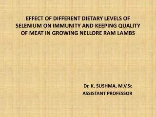
Cont….. Composition of Blood • Monocytes: – Compose 4–8% of WBCs • The largest circulating leukocytes • Size: 12 – 20 µm in diameter • Nucleus : kidney shaped • Transform into macrophages • Phagocytic cells
Monocytes Eosinophils
Lymphocytes Basophils
Cont….. Composition of Blood Platelets: • Structure • Small disc shaped, without nucleus having 2-4 µm diameter; originate in bone marrow from giant cell megakaryocyte • Alpha granules contain several • clotting factors • platelet derived growth factors – Dense granules contain ADP, ATP, calcium ions, serotonin and fibrin stabilizing factor. • Function • Involved in stopping bleeding when a blood vessel is damaged; Process is called Hemostasis
Mechanism of Hemostasis • When a blood vessel is damaged, a number of physiological mechanisms are activated that promote the process of Hemostasis • Mechanisms of Hemostasis: -Vasoconstriction - Platelet plug formation - Clot formation
Steps in Vascular Damage and Clotting Responses • Vasoconstriction: contraction of smooth muscle in the walls of arteriole to reduce blood flow. • Platelet plug formation: 1. Platelet adhesion: Platelets contact and stick to free collagen fibers of the damaged blood vessel 2. Platelet release reaction: Activated platelets extend many projections that enable them to contact and interact with one another. They then liberate their granules.
Steps in Vascular Damage and Clotting Responses The liberated ADP and thromboxane A2 help to activate other platelets. Serotonin and thromboxane A2 act as vasoconstrictors helping to decrease blood flow. 3. Platelet aggregation: Liberated ADP makes new platelets sticky; these newly-recruited and activated platelets adhere to the originally-activated platelets. The process starts the formation of a platelet plug.
Steps in Vascular Damage and Clotting Responses • Plate aggregation is regulated by two chemical mediators (eicosanoids) i-e • Thromboxane A2 (TXA2) Primary source is damaged blood vessels that stimulate platelet aggregation. • Prostacyclin (PGI2) Primary source of prostacyclin is intact blood vessels that inhibits platelet aggregation.
Steps in Vascular Damage and Clotting Responses • In the absence of vessel damage, platelets are repelled from each other and from the endothelial lining of vessels • The repulsion of platelets from an intact endothelium is believed to be due to prostacyclin produced with in the intact endothelium
Damage to the endothelium of vessels leads to secretion of Von Willebrand factor by endothelial cells • This factor initiate the adhesion of platelets to the damaged vessel • When the platelets adhere to the vessel, platelets release ADP ( Adenosin Diphosphate ), serotonin and Thromboxane A2
Serotonin and Thromboxane A2 stimulate vasoconstriction, which helps to decrease blood flow to the injured blood vessel • Phospholipids that are exposed on the platelet membrane participate in the activation of clotting factors • The release of ADP and Thromboxane A2 from platelets make other platelets also sticky • This produces a platelet plug
Clotting Factors: Formation of fibrin Clot formation • The platelet plug is strengthen by a mesh work of insoluble protein fibers known as fibrin then the platelet pug is converted into firm clot • Blood clots contain platelets and fibrin, and they usually contain trapped red blood cells that give the clot a red color • Finally, contraction of the platelet mass in the process of clot retraction forms more effective and more compact plug
The conversion of fibrinogen in to fibrin may occur via 2 pathways Intrinsic pathway Extrinsic pathway • By both these pathways clotting factors get activated • This leads to activation of inactive enzyme Prothrombin to active enzyme Thrombin • Thrombin converts the soluble protein fibrinogen in to insoluble protein fibrinwhich form the mesh work supporting platelet plug.
Dissolution of clots • As damaged blood vessel wall is repaired factor XII promotes conversion of Kallikrein from inactive form to active form • This Kallikrein convert inactive Plasminogen in to active Plasmin • Plasmin is an enzyme that digests fibrin into split products
The Blood Clotting Cascade • Extrinsic Pathway (Fast acting) 1. Tissue Factor (TF) or Thromboplastin is released by tissue cells outside of the damaged vessel. 2. TF begins a chemical reaction pathway that activates Thrombokinase (F10). F10 combines with Proaccelerin (F5) to form the enzyme Prothrombinase.
The Blood Clotting Cascade • Intrinsic Pathway (slow acting) Activated by factors within the blood or vessels Antihemophilic factor D or Hageman factor (F12) is activated by contact with collagen fibers. F12 starts a chemical cascade that ultimately activates F10 or Thrombokinase. F10 combines with Proaccelerin (F5) to form the enzyme Prothrombinase.
The Blood Clotting Cascade • The Common Pathway Prothrombinase catalyzes the conversion of Prothrombin (F2) to Thrombin. Thrombin converts the soluble plasma protein fibrinogen in the insoluble protein fibrin (loose threads). Thrombin also activates Fibrin Stabilizing Factor (F13) which converts the loose threads into stable threads.
The Problems with Clotting Cascade • Hemophilia A: Deficiency of Factor VIII accounts for 85% cases. • Almost exclusively in males. Females are usually carriers • caused by a gene mutation on the “X” chromosome. Occurs in about 1/10,000 male births • Other Hemophilias account for another 15% • Hemophilia B (Factor IX) • Hemophilia C (Factor XI) • Hemophilia D (Factor XII)
The Problems with Platelets and Abnormal Clotting • Thrombocytopenia: Abnormally low levels of platelets. Usually below 50,000/µl of blood. • In many cases, specific antibodies are produced against platelets destroying them • Thrombus: Abnormal clot that develops in a blood vessel. • Embolus:Free thrombic clots carried in the blood that usually get caught in arterioles in the brain, kidney, and lungs.
White Blood Cell Disorders Pathological Terms White Blood Cell Disorders Granulocytosis Leukemia • Neoplastic disorder in which there is an excessive increase in white blood cells • Abnormal increase of granulocytes in the bloodstream. Commonly seen during times of infection Multiple Myeloma • Malignant tumor of the bone marrow