Download
1 / 50
500 likes | 623 Views
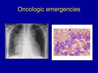
Oncologic challenges in the ED. (besides not getting the old chart from TBCC) Grand Rounds Gord McNeil. 6 Cases Approach Management Calgary perspective. Case 1.

E N D
Oncologic challenges in the ED (besides not getting the old chart from TBCC) Grand Rounds Gord McNeil
6 Cases • Approach • Management • Calgary perspective
Case 1 • 52 year old female with breast cancer presents to the ED with mid back discomfort, progressive weakness of left leg X 1 week and today urinary incontinence • Recent radiation at TBCC (no old chart available)
Approach • Physical • T=37.3 Hr=92, RR=14, BP=172/89 • Decreased sensation left abdominal wall and right lower leg • Decreased power at right knee and ankle • Labs • Hg=109, Plts =302, WBC =6.8, normal lytes and INR.
Differential diagnosis • Epidural abscess • Epidural hematoma • Metastatic spinal cord compression • Routine causes of back pain
Treatment • Dexamethasone IV 10mg prior to MRI, then 4-8 mg q6-8hours • Emergent MRI of entire spine (because pt can have synchronous, multifocal, asymptomatic MSCC.
Treatment • Call Spine service • Decompression of spinal cord is the key to salvage of function • Patchell et al2 in 2005 - radiation for 10 days and decompressive surgery within 24 hours improved outcomes of ambulation, continence and functional abilities from 84% compared to radiation alone for 57%
Metastatic spinal cord compression • Causes • breast(30%), • lung (15%) • prostate (15%) • Other • Sites • thoracic, then lumbar then cervical
MSCC - causes • Expansion of vertebral bone metastasis into epidural space causing cord compression – radiation helps • Neural foramina extension by a paraspinal mass. – radiation helps • Destruction of vertebral cortical bone -requires surgical intervention.
Prognosis • Start of onset of symptoms: Onset: 1-7 days8-14days>14 days Ambulate: 35% 55% 86% (1) • Faster onset = worse prognosis • Start of therapy: dexamethasone and time to surgery • Favorable histology - radiosensitive tumors
Treatment • Radiation only arrests the progression of nonradiosensitive tumors and does not stabilize the spine • Surgery allows immediate cord decompression whereas radiotherapy typically takes several days to weeks.
Calgary perspective • Radiation oncology – Dr. Elizabeth Yan • Radiation did have an important initial role prior to 2005. Now acute surgical decompression and post op radiation is the standard of care.
Calgary perspective • Case scenarios • Highly suspicious for occult CA and back pain then plain films and MRI – no steroids • Known CA and back pain without neuro deficit then MRI, steroids and radiation oncology • Known CA with neuro deficit, then steroids, MRI and spine service
Case 2 • 48 yr old male presents to ED with large hemoptysis X 2 • Recently treated at TBCC for lung CA (old chart not available) • HR =129, RR=32, sat=90% 5L, BP=167/96
Approach • Mobilize team early • Pulmonary • DI/ IR • ICU • Thoracics
Approach • Stabilize • Unstable airway • ETT – large size to faciliate bronchoscope • Not the panacea • Pulmonary toilet – very important • Selective placement of ETT
Approach • Stabilize • CXR –localizes bleeding • Patient position – bleeding side down • Blood products/ fluids prn
Approach • Imaging • CT scan can be done if pt not intubated and has stable airway prior to interventional radiology for bronchial artery emobilization • If ETT then often bronch before IR to localize bleeding
Approach • Hemoglobin not important • patients die of hypoxia not anemia • not like GI bleed
Causes • Friable endobronchial tumors • tumor eroding into a small intrapleural vessel • tumour eroding in to one of the major vessels of the thorax. • Large vessels bleeds = death
Calgary perspective • Dr. Alain Tremblay • One of the few indications for stat call for pulmonary in the middle if the night – involve pulmonary early • Mobilize CT and Interventional radiology early • Supportive management essential
Case 3 • 73 yr old male with thyroid cancer c/o increased secretions, stridor and SOB. • HR = 112, RR=36, BP=178/102, sat=91%on NRB
Approach • Stabilize • O2, suctioning of secretions and allowing patient to sit up • Labs, CXR
Why is it happening? • Usually a subacute process unless an already marginal airway is suddenly compromised by an acute infection, bleeding or the patient’s inability to handle secretions. • Thyroid and esophageal carcinomas may compress the trachea by invading the surrounding soft tissue • Can occur from scarring from prolonged intubation or from radiation therapy
Treatment Consultant • Pulmonary – Rigid scope for endobronchial stenting or laser abalation • Steroids – not helpful (only if known lymphoma)
Calgary perspective • Needs rigid scope • Drs Tremblay and Michaud only 2 pulmonologist in Calgary who do rigid scope (Some thoracic surgeons do as well) • Can call pulmonary at any site and then can help management patient and arrange for rigid scope
Case 4 • 86 yr old female with metastatic lung CA with progressive SOBOE over last 2 weeks, now SOB at rest. • Nonproductive cough, no fever. • HR =92, RR=24, BP 164/92 Sat=94% on 2L
Approach • Stabilize • Labs • CXR • Pleurocentesis
Why is it happening ? • Most common from lung, breast, ovary and lymphoma • Pleural seeding by neoplastic cells increases capillary permeability and produces an exudative effusion • Direct erosion into a blood vessel can cause an abrupt hemorrhagic effusion
Calgary perspective • Dyspnea clinic • Run by Dr. Trembaly and Dr. Michaud • Refer if known CA with symptomatic effusion or if highly suspicious for cancer • Don’t necessarily need tissue diagnosis
Dyspnea Clinic • Tap in ED, send referral. Appt usually in 2 weeks • Clinic places pleurodex catheter and have home care drain it off as necessary • If tapped in ED and return prior to appt, may need admission to pulmonary • Clinic number -521 3511 – Pat Barkley
Case 5 • 64 yr old female with metastatic breast CA to liver “flu –like” symptoms, N/V, lethargy, weakness X 2 weeks • HR =110, RR=16, BP=100/56, Sat =84% RA • GCS = 13, no focal deficit, clinically “dry”
Approach • Labs • Hg =112, WBC =9.4 Plts =186 • Glc = 7.5, Na =132, K = 3.5 • Creatinine =364 (new) • Calcium= 3.64 albumin =29 • Management
Treatment • Measure ionized calcium • ABG • Corrected calcium = measured calcium + (0.02 X(40 – measured albumin) • Lower the albumin and the corrected calcium goes up
Treatment • Replace volume first • Sodium inhibits reabsorption of calcium • Need urine output – 100cc/hr • After euvolemic, then lasix with volume maintenance • Follow K and Mg closely
Causes of hypercalcemia in malignancy • One of the most common complications of cancer - 10-20% • MC caused by breast, lung, renal and cholangiocarcioma and multiple myeloma and lymphoma • Mobilization of bone calcium more rapidly than it can be cleared by the kidneys • Secretion of parathyroid hormone • Presence of bone mets that cause local destruction
Case 6 • 62 yr old male with CML with a recent exacerbation of COPD put on prednisone and levaquin • Acute onset of flank pain then new tonic clonic seizure x 3 minutes • Hr =48, RR =28, BP = 88/52, sat =94%NRB, T=37.6, C/S=6.8
Approach • Stabilize • Labs • Hg = 109, WBC =38, plts=201 • K = 6.8, Na = 132, glc = 6.9 • Cr= 342, urea =32 • Calcium = 1.87, Phosphate = 2.78, albumin =38 • Diagnosis ?
Tumor Lysis Syndrome • Hyperkalemia • Hyperphosphatemia • Hypocalcemia • Renal failure • Renal colic
Tumour lysis syndrome - causes • Large burden of tumor is rapidly and acutely destroyed causes outpouring of potassium, nucleic acids and phosphates. • Sudden build up of electrolytes • MC seen with lymphoma and leukemia, but can also occur with solid organ tumors • Usually within 6 hours to 6 days after the initiation of therapy • Can occur with the administration of corticosteroids to a susceptible patient
Symptoms of hyperkalemia • - weakness and altered MS and arrthymias • Hyperphsophatemia • Causes acute precipitation of calcium in the kidneys and tissues leading to…. • Symptoms of hypocalcemia • carpopedal spasm and seizures • Renal failure • secondary to increased uric acid levels producing renal tubular necrosis • Symptoms of renal colic • secondary to increased uric acid levels producing renal tubular necrosis
Treatment -Tumor lysis syndrome • Aggressive hydration if urine output exists • Alkalinization of urine to pH 7 (can worsen hypocalcemia) • Correct electrolytes and follow closely • Lasix • Allopurinol – 600- 900mg loading dose • Hemodialysis
Rad onc, Med onc, no onc…who goes where? • Radiation therapy • Patient with active radiation – usually gets s/e 2 weeks after starting radiation until 2 weeks after completing radiation – eg diarrhea • Medical oncology • Patient with chemo within the last month • Usually febrile neutropenia at 5 days • No oncology No tissue diagnosis?? – hospitalist
References • 1) pg 508 - hematology/oncology clinics of north america • 2 pg 521 – radiation oncology emergencies
1) MSCC • 2) Hemoptysis • 3) Malignant effusion • 4) hypercalcemia • 5) Tumor lysis syndrome • 6) Airway compromise • Hyperviscosoity syndrome • SVC syndrome • SIADH