Download
1 / 153
1.53k likes | 1.56k Views
Explore the fascinating journey of prokaryotes from the emergence of the first cells to the diversification of life forms, uncovering the conditions that allowed for the origin of life on Earth and the synthesis of organic molecules. Discover the significance of RNA in the early genetic material and fossil evidence of ancient prokaryotic life.

E N D
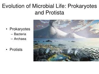
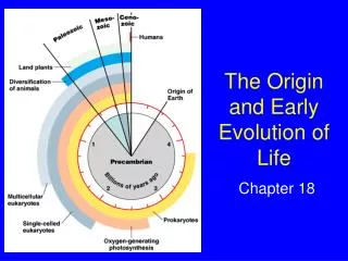
Earth formed 4.6 billion years ago The oldest fossil organisms are prokaryotes dating back to 3.5 billion years ago Prokaryotes are single-celled organisms in the domains Bacteria and Archaea Some of the earliest prokaryotic cells lived in dense mats that resembled stepping stones Overview: The First Cells
Prokaryotes are the most abundant organisms on Earth There are more in a handful of fertile soil than the number of people who have ever lived Prokaryotes thrive almost everywhere, including places too acidic, salty, cold, or hot for most other organisms Some prokaryotes colonize the bodies of other organisms
Concept 24.1: Conditions on early Earth made the origin of life possible Chemical and physical processes on early Earth may have produced very simple cells through a sequence of stages Abiotic synthesis of small organic molecules Joining of these small molecules into macromolecules Packaging of molecules into protocells, membrane-bound droplets that maintain a consistent internal chemistry Origin of self-replicating molecules
Synthesis of Organic Compounds on Early Earth Earth’s early atmosphere likely contained water vapor and chemicals released by volcanic eruptions (nitrogen, nitrogen oxides, carbon dioxide, methane, ammonia, and hydrogen) As Earth cooled, water vapor condensed into oceans, and most of the hydrogen escaped into space
In the 1920s, A. I. Oparin and J. B. S. Haldane hypothesized that the early atmosphere was a reducing environment In 1953, Stanley Miller and Harold Urey conducted lab experiments that showed that the abiotic synthesis of organic molecules in a reducing atmosphere is possible
However, the evidence is not yet convincing that the early atmosphere was in fact reducing Instead of forming in the atmosphere, the first organic compounds may have been synthesized near volcanoes or deep-sea vents Miller-Urey-type experiments demonstrate that organic molecules could have formed with various possible atmospheres Organic molecules have also been found in meteorites Video: Hydrothermal Vent Video: Tubeworms
Figure 24.3 20 200 Number of amino acids Mass of amino acids (mg) 10 100 0 0 1953 2008 1953 2008
Abiotic Synthesis of Macromolecules RNA monomers have been produced spontaneously from simple molecules Small organic molecules polymerize when they are concentrated on hot sand, clay, or rock
Protocells Replication and metabolism are key properties of life and may have appeared together Protocells may have been fluid-filled vesicles with a membrane-like structure In water, lipids and other organic molecules can spontaneously form vesicles with a lipid bilayer
Adding clay can increase the rate of vesicle formation Vesicles exhibit simple reproduction and metabolism and maintain an internal chemical environment
Figure 24.4 0.4 Precursor molecules plus montmorillonite clay Relative turbidity, an index of vesicle number 0.2 Precursor molecules only 0 40 0 60 20 Time (minutes) (a) Self-assembly 1 m Vesicle boundary 20 m (b) Reproduction (c) Absorption of RNA
Figure 24.4a 0.4 Precursor molecules plus montmorillonite clay Relative turbidity, an index of vesicle number 0.2 Precursor molecules only 0 40 60 0 20 Time (minutes) (a) Self-assembly
Figure 24.4b 20 m (b) Reproduction
Figure 24.4c 1 m Vesicle boundary (c) Absorption of RNA
Self-Replicating RNA The first genetic material was probably RNA, not DNA RNA molecules called ribozymes have been found to catalyze many different reactions For example, ribozymes can make complementary copies of short stretches of RNA
Natural selection has produced self-replicating RNA molecules RNA molecules that were more stable or replicated more quickly would have left the most descendant RNA molecules The early genetic material might have formed an “RNA world”
Vesicles with RNA capable of replication would have been protocells RNA could have provided the template for DNA, a more stable genetic material
Fossil Evidence of Early Life Many of the oldest fossils are stromatolites, layered rocks that formed from the activities of prokaryotes up to 3.5 billion years ago Ancient fossils of individual prokaryotic cells have also been discovered For example, fossilized prokaryotic cells have been found in 3.4-billion-year-old rocks from Australia
Figure 24.5 30 m 5 cm 10 m Stromatolites Nonphotosynthetic bacteria Possible earliest appearance in fossil record Cyanobacteria 1 4 3 2 0 Time (billions of years ago)
Figure 24.5a Stromatolites Nonphotosynthetic bacteria Possible earliest appearance in fossil record Cyanobacteria 1 4 3 2 0 Time (billions of years ago)
Figure 24.5b 30 m 3-billion-year-old fossil of a cluster of nonphotosynthetic prokaryote cells
Figure 24.5c 5 cm 1.1-billion-year-old fossilized stromatolite
Figure 24.5d 10 m 1.5-billion-year-old fossil of a cyanobacterium
The cyanobacteria that form stromatolites were the main photosynthetic organisms for over a billion years Early cyanobacteria began the release of oxygen into Earth’s atmosphere Surviving prokaryote lineages either avoided or adapted to the newly aerobic environment
Concept 24.2: Diverse structural and metabolic adaptations have evolved in prokaryotes Most prokaryotes are unicellular, although some species form colonies Most prokaryotic cells have diameters of 0.5–5 µm, much smaller than the 10–100 µm diameter of many eukaryotic cells Prokaryotic cells have a variety of shapes The three most common shapes are spheres (cocci), rods (bacilli), and spirals
Figure 24.6 1 m 1 m 3 m (a) Spherical (b) Rod-shaped (c) Spiral
Figure 24.6a 1 m (a) Spherical
Figure 24.6b 1 m (b) Rod-shaped
Figure 24.6c 3 m (c) Spiral
Cell-Surface Structures A key feature of nearly all prokaryotic cells is their cell wall, which maintains cell shape, protects the cell, and prevents it from bursting in a hypotonic environment Eukaryote cell walls are made of cellulose or chitin Bacterial cell walls contain peptidoglycan, a network of modified sugars cross-linked by polypeptides
Archaeal cell walls contain polysaccharides and proteins but lack peptidoglycan Scientists use the Gram stain to classify bacteria by cell wall composition Gram-positive bacteria have simpler walls with a large amount of peptidoglycan Gram-negative bacteria have less peptidoglycan and an outer membrane that can be toxic
Figure 24.7 (a) Gram-positive bacteria (b) Gram-negative bacteria Carbohydrate portion of lipopolysaccharide Peptido- glycan layer Outer membrane Cell wall Cell wall Peptidoglycan layer Plasma membrane Plasma membrane Gram-positive bacteria Gram-negative bacteria 10 m
Figure 24.7a (a) Gram-positive bacteria Peptido- glycan layer Cell wall Plasma membrane
Figure 24.7b (b) Gram-negative bacteria Carbohydrate portion of lipopolysaccharide Outer membrane Cell wall Peptidoglycan layer Plasma membrane
Figure 24.7c Gram-negative bacteria Gram-positive bacteria 10 m
Many antibiotics target peptidoglycan and damage bacterial cell walls Gram-negative bacteria are more likely to be antibiotic resistant A polysaccharide or protein layer called a capsule covers many prokaryotes
Figure 24.8 Bacterial capsule Bacterial cell wall Tonsil cell 200 nm
Some bacteria develop resistant cells called endospores when they lack an essential nutrient Other bacteria have fimbriae, which allow them to stick to their substrate or other individuals in a colony Pili (or sex pili) are longer than fimbriae and allow prokaryotes to exchange DNA
Figure 24.9 Fimbriae 1 m
Motility In a heterogeneous environment, many bacteria exhibit taxis, the ability to move toward or away from a stimulus Chemotaxis is the movement toward or away from a chemical stimulus
Most motile bacteria propel themselves by flagella scattered about the surface or concentrated at one or both ends Flagella of bacteria, archaea, and eukaryotes are composed of different proteins and likely evolved independently
Figure 24.10 Flagellum 20 nm Filament Hook Motor Cell wall Peptidoglycan layer Plasma membrane Rod
Figure 24.10a 20 nm Hook Motor
Evolutionary Origins of Bacterial Flagella Bacterial flagella are composed of a motor, hook, and filament Many of the flagella’s proteins are modified versions of proteins that perform other tasks in bacteria Flagella likely evolved as existing proteins were added to an ancestral secretory system This is an example of exaptation, where existing structures take on new functions through descent with modification
Internal Organization and DNA Prokaryotic cells usually lack complex compartmentalization Some prokaryotes do have specialized membranes that perform metabolic functions These are usually infoldings of the plasma membrane
Figure 24.11 1 m 0.2 m Respiratory membrane Thylakoid membranes (a) Aerobic prokaryote (b) Photosynthetic prokaryote