Download
1 / 30
300 likes | 350 Views
Learn about epithelial, connective, muscle, and nervous tissues and their functions. Explore the characteristics and locations of each tissue type.

E N D
Ch 4: Tissue • Groups of cells that are similar in structure and perform a common or related functions are called TISSUE • The study of tissue, or histology, complements the study of gross anatomy. Together they provide the structural basis for understanding organ physiology.
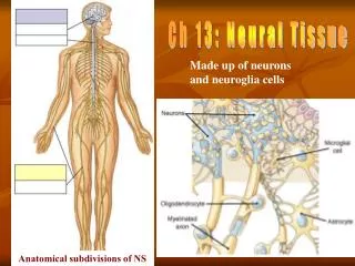
Tissues • The tissues of the human body include four major types: general function • Epithelial: covering • Connective: support • Muscle: movement • Nervous: control
EPITHELIAL TISSUES • Epithelial Tissue: is a sheet of cells that covers a body surface or lines a body cavity • Nearly all substances received or given off by the body must pass through an epithelium layer.
EPITHELIAL TISSUES • Epithelial Tissue lacks of blood vessels, contains little intercellular material and are continually being replaced • They function in protection, secretion, absorption, filtration, excretion, and sensory reception.
Epithelium Tissue has many characteristics that separate it from other tissue types ET has Polarity- which means it has an apical surface (near the top) and a basal surface (near the bottom) Specialized Contacts Supported by connective tissue. EPITHELIAL TISSUES
. EPITHELIAL TISSUES • Each Epithelium Tissue is given two names. • The first name indicates the number of cell layers present. • Simple- one layer • Stratified- more than one layer • The second describes the shape of its cells • Squamous, cuboidal, and columnar.
. EPITHELIAL TISSUES
Simple Squamous Epithelium • This tissue consists of a single layer of thin, flattened cells through which substances can pass easily. So delicate they can easily be damaged. • Common site of diffusion and osmosis. Its functions in the exchange of gases in the lungs • Lines the air sacs of the lungs (gas exchange), forms the walls of the capillaries, lines the insides of blood vessels and lymph vessels. • Covers the membrane that line body cavities. • EX: Cheek Cells
Simple Cuboidal Epithelium • This tissue consists of a single layer of cube-shaped cells. Usually have a centrally located spherical nuclei • It carries on secretion and absorption. Secretes glandular products. • Covers the ovaries, lines the kidneys, tubules and ducts of certain glands like pancreas and the liver.
Simple Columnar Epithelium • The tissue is composed of a single layer of elongated cells whose nuclei are usually at about the same level, near the basement membrane. • Some have cilia some do not • Secretes and absorbs…this tissue is thick!! • This tissue lines uterus and portions of the digestive tract like small/large intestines and stomach.
Pseudostratified Columnar Epithelium • They appear stratified but are NOT, They appear to have two or more nuclei but they each reach the basement level. • Goblet cells scattered throughout the tissue that secret mucus, which the cilia sweep away. • It lines tubes of the respiratory system. The mucus and cilia created by this tissue trap the dust and microorganisms that enter the airway.
Pseudostratified Columnar Epithelium • “C” the nuclei appear at various levels giving it the stratified appearance • “B” the cilia can easily be seen
Stratified Squamous Epithelium • Named for the shape of the cells,…consists of many layers, relatively thick. Cells nearer the surface are flat where as the deeper are cuboidal and columnar. • Tissue can contain Keratin, which is a protein that accumulates and protect the underlying tissue. • Epidermis-outer most layer of the skin • Also lines the skin and lines the oral cavity, throat, vagina, and anal canal.
Stratified Squamous Epithelium • Observe that the surface cells (at B) are flattened (are squamous).
Stratified Cuboidal Epithelium • This tissue is composed of two or thee layers of cube-shaped cells. • It lines the larger ducts of the mammary glands sweat glands, salivary glands, and pancreas. Forms the lining of ovarian follicles and seminiferous tubules • It functions in protection.
Transitional Epithelium • This tissue is specialized to change in response to tension.. • Transition epithelium is unstretched and consists of many layers when the organs wall contract the tissue stretches and appears thinner when the organ is distended. • Forms the lining of the urinary bladder and lines the ureters and part of the urethra.