Download
1 / 17
190 likes | 607 Views
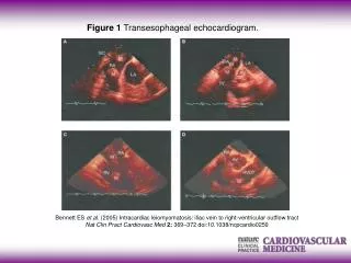
Transesophageal Echocardiography Assisted Amplatzer Occlusion of PDA. Bill Lee Trapper Belk Acc# 109608. TEE PDA. Trapper Belk (ID# 146088) 9 Mo, MC Bloodhound PDA Dx’d at UGA – referred for occlusion. Cardiomegaly, L atriomegaly Interstitial pattern – poss. CHF.

E N D
Transesophageal Echocardiography Assisted Amplatzer Occlusion of PDA Bill Lee Trapper Belk Acc# 109608
TEE PDA • Trapper Belk (ID# 146088) • 9 Mo, MC Bloodhound • PDA Dx’d at UGA – referred for occlusion
Cardiomegaly, L atriomegaly • Interstitial pattern – poss. CHF
Prox. descending aorta, MPA enlargement • Pulm. Vasc. enlargement
≈ $30,000 probe • Inner knob – flexes and extends probe • Outer knob – pans tranducer forward and backward • Side to side panning via manual rotation
Position probe on prox. desc. aorta • Use inner knob to improve contact b/w probe and ventral esoph. • Use outer knob and manual rotation to optimize
Transducer direction arrow • Marker toward head • Watch probe temp! – esophageal burn/perf. major complication of procedure
Optimize PDA • Measure diameter of narrowest (distal) portion of ductus for Amplatzer sizing