Download
1 / 84
840 likes | 974 Views
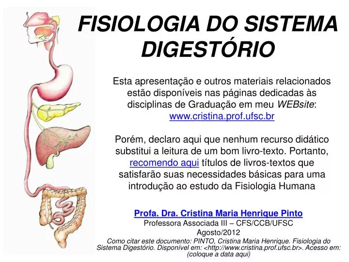
FISIOLOGIA DO SISTEMA DIGESTÓRIO. Esta apresentação e outros materiais relacionados estão disponíveis nas páginas dedicadas às disciplinas de Graduação em meu WEBsite : www.cristina.prof.ufsc.br

E N D
FISIOLOGIA DO SISTEMA DIGESTÓRIO Esta apresentação e outros materiais relacionados estão disponíveis nas páginas dedicadas às disciplinas de Graduação em meu WEBsite: www.cristina.prof.ufsc.br Porém, declaro aqui que nenhum recurso didático substitui a leitura de um bom livro-texto. Portanto, recomendo aqui títulos de livros-textos que satisfarão suas necessidades básicas para uma introdução ao estudo da Fisiologia Humana Profa. Dra. Cristina Maria Henrique Pinto Professora Associada III – CFS/CCB/UFSC Agosto/2012 Como citar este documento: PINTO, Cristina Maria Henrique. Fisiologia do Sistema Digestório. Disponível em: <http://www.cristina.prof.ufsc.br>. Acesso em: (coloque a data aqui)
FISIOLOGIA DO SISTEMA DIGESTÓRIO Títulos das apresentações disponíveis: • Introdução ao estudo do sistema digestório (SD) • Movimentos observados no trato gastrointestinal (TGI) • Secreções do TGI • Digestão e absorção dos principais nutrientes de uma dieta ideal
FISIOLOGIA DO SISTEMA DIGESTÓRIO Títulos desta apresentação: • Secreções do TGI • Introdução. Conceitos gerais • Secreção salivar: • Componentes, funções e regulação da secreção. • Secreção gástrica: • Componentes, funções e regulação da secreção. • Secreções pancreática, hepática e intestinais: • Componentes, funções e regulação da secreção.
FISIOLOGIA DO SISTEMA DIGESTÓRIO Títulos desta apresentação: • Secreções do TGI • Introdução. Conceitos gerais • Secreção salivar: • Componentes, funções e regulação da secreção. • Secreção gástrica: • Componentes, funções e regulação da secreção. • Secreções intestinais: • Componentes, funções e regulação da secreção.
Resumo das secreções exócrinas do TGI Digestive System (Vander, Sherman & Luciano, 2002, McGraw-Hilll)– WEBsite original enquantodisponível: http://www.biocourse.com
Tipos de secreções do TGI NEURÓCRINA (neurotransmissores e/ou neuromoduladores) ENDÓCRINA (hormônios) EXÓCRINA (mucosa, serosa e/ou hidroeleletrolítica)
FISIOLOGIA DO SISTEMA DIGESTÓRIO Títulos desta apresentação: • Secreções do TGI • Introdução. Conceitos gerais • Secreção salivar: • Componentes, funções e regulação da secreção. • Secreção gástrica: • Componentes, funções e regulação da secreção. • Secreções intestinais: • Componentes, funções e regulação da secreção.
As glândulas salivares There are several pairs of salivary glands in different locations: a major pair in front of the ears (parotid glands); two major pair on the floor of the mouth (sublingual and submaxillary glands); and several minor pairs within the lips, cheeks, and tongue. http://www.nlm.nih.gov/medlineplus/ency/imagepages/9654.htm
Histologia das glândulas salivares • Células acinares(serosa, mucosa ou sero-mucosa) • Células ductais(intercalar, estriado e excretor) • Células mioepiteliaislocalizadas entre a membrana basal e as células acinares. Modificado de Junqueira e Carneiro, 2001
Composição da saliva Água (98-99%), Produtos Inorgânicos e Orgânicos PRODUTOS ORGÂNICOS: Compostos por proteínas salivares de 4 tipos: • P. Enzimáticas: • AMILASE: Inicia a degradação do amido e do glicogênio, mas tem um papel pequeno porque se inativa rapidamente pelo fluxo digestivo. • LACTOPEROXIDASE: Ação antibacteriana destrói os microorganismos ao catalizar o peróxido de oxigênio. • LISOZIMA : Ação antibacteriana, inibe o crescimento bacteriano, reduz a incorporação de glicose e produz ácido láctico. • P. ricas en prolina: • MUCINAS: Capacidade de formar uma pseudomembrana sobre superfícies finas e duras, tem uma função protetora. São proteínas ácidas ricas em prolina. • P. Aromáticas: • GUSTINA, que agudiza o gosto. • ESTATERINA, que produz remineralização e evita a precipitação ou cristalização de sais de fosfato de cálcio supersaturado nos ductos salivares HISTATINA, que liga-se à hidroxiapatita; idem acima • LACTOFERRINA, intervém no retardo do crescimento bacteriano. • ALBUMINA, que produz compostos aromáticos. • Imunoglobulinas (IgA). • PRODUTOS INORGÂNICOS: Cálcio, flúor, Sódio, Potássio, Bicarbonato, Fosfato, Cloro, Magnésio. http://html.rincondelvago.com/desordenes-salivales_saliva-y-medio-bucal.html e http://nossodentista.com/saliva.htm
A secreção salivar É produzida 24 h/dia graças ao tônus Parassimpático, mesmo durante o sono...
Secreção das glândulas salivares Representação esquemática do modelo de secreção salivar em dois estágios. ácino A ritmos máximos de secreção, as glândulas salivares podem secretar até 1 ml/min por grama de tecido, isto é, o próprio peso por minuto! Ducto impermeável à água
A secreção serosa salivar: alfa-amilase (ptialina) Amilopectina (amido) de batata
O médico russo Ivan Petrovich Pavlov (1849 - 1936) percebeu que a apresentação de alimento desencadeava, em cães famintos, um reflexo natural de salivação. A associação sistemática entre a apresentação de alimento e o barulho de uma campainha, provocava, depois de um certo tempo, o reflexo condicionado, ou seja, apenas o som da campainha era capaz de desencadear de salivação no cão faminto. . http://nobelprize.org/medicine/laureates/1904/pavlov-bio.html
Reflexos incondicionados • São aqueles que estimulam a salivação sem que haja o aprendizado (p. ex., apresentação de comida a um indivíduo faminto). O médico russo Ivan Petrovich Pavlov (1849 - 1936) percebeu que a apresentação de alimento desencadeava, em cães famintos, um reflexo natural de salivação. A associação sistemática entre a apresentação de alimento e o barulho de uma campainha, provoca, depois de um certo tempo, o reflexo condicionado, ou seja, apenas o som da campainha é capaz de desencadear de salivação no cão faminto. . http://nobelprize.org/medicine/laureates/1904/pavlov-bio.html
O médico russo Ivan Petrovich Pavlov (1849 - 1936) percebeu que a apresentação de alimento desencadeava, em cães famintos, um reflexo natural de salivação. A associação sistemática entre a apresentação de alimento e o barulho de uma campainha, provocava, depois de um certo tempo, o reflexo condicionado, ou seja, apenas o som da campainha era capaz de desencadear de salivação no cão faminto. . http://nobelprize.org/medicine/laureates/1904/pavlov-bio.html
Reflexos condicionados Reflexos condicionados • São os que necessitam de experiência prévia, repetitiva e associativa entre alimentação e olfação/visão. O médico russo Ivan Petrovich Pavlov (1849 - 1936) percebeu que a apresentação de alimento desencadeava, em cães famintos, um reflexo natural de salivação. A associação sistemática entre a apresentação de alimento e o barulho de uma campainha, provocava, depois de um certo tempo, o reflexo condicionado, ou seja, apenas o som da campainha era capaz de desencadear de salivação no cão faminto. . Pavlov recebeu o Prêmio Nobel em 1904 de Fisiologia e Medicina, por suas pesquisas. http://nobelprize.org/medicine/laureates/1904/pavlov-bio.html
Reflexos condicionados • São os que necessitam aprendizado prévio e repetitivo, como a olfação e a visão. • Ex: uma criança lactente não reage (salivando) como um adulto.
Regulação da secreção salivar Produção e secreção de saliva 24 horas/dia Tônus PS
Regulação da secreção salivar (+) ou (-) PS: Sistema Nervoso Parassimpático; SP: Sistema Nervoso Simpático; GCS: gânglio cervival superior.
Regulação da secreção salivar extraído, enquanto disponível, de Pocock & Richards, : http://www.oup.com/uk/orc/bin/9780198568780/
“Centros” superiores “Centros” superiores Tronco encefálico “Centro” da mastigação “Centro” da deglutição “Centro” da salivação estímulos mastigatórios estímulos gustativos distensão gástrica Os principais componentes envolvidos na ativação neural das glândulas salivares olfação Início da salivação por reflexos incondicionados Tronco encefálico “Centro” da mastigação “Centro” da deglutição “Centro” da salivação N. V estímulos mastigatórios N. VII, IX, X estímulos gustativos ramos PS N. VII N. IX I-OLFATÓRIO II-ÓPTICO III-OCULOMOTOR IV-TROCLEAR V-TRIGÊMEO VI-ABDUCENTE VII-FACIAL VIII-VESTÍBULO- COCLEAR IX-GLOSSOFARÍNGEO X-VAGO XI-ACESSÓRIO XII-HIPOGLOSSO glândulas submandibulares e sublinguais glândulas parótidas ramos SP gânglio cervical superior Pedersen et al., 2002: Saliva and gastrointestinal functions of taste, mastication, swallowing and digestion.Oral Diseases 8 (3), 117-129, 2002. Caso não seja possível o acesso, peça cópia à Profa. Cristina segmento superior torácico da medula espinhal
“Centros” superiores “Centros” superiores Tronco encefálico “Centro” da mastigação “Centro” da deglutição “Centro” da salivação N. V estímulos mastigatórios N. VII, IX, X estímulos gustativos distensão gástrica Os principais componentes envolvidos na ativação neural das glândulas salivares visão, olfação e pensamento Tronco encefálico Início da salivação por reflexos condicionados: A visão, olfação e o pensamento podem levar à formação de alguma saliva, dependendo do estado motivacional. Os “núcleos salivatórios” também recebem aferências de outras regiões do SNC que podem resultar em efeitos estimulatórios ou inibitórios sobre a salivação, dependendo, por exemplo, do estado emocional. “Centro” da mastigação I-OLFATÓRIO II-ÓPTICO III-OCULOMOTOR IV-TROCLEAR V-TRIGÊMEO VI-ABDUCENTE VII-FACIAL VIII-VESTÍBULO- COCLEAR IX-GLOSSOFARÍNGEO X-VAGO XI-ACESSÓRIO XII-HIPOGLOSSO “Centro” da deglutição “Centro” da salivação N. V estímulos mastigatórios N. VII, IX, X estímulos gustativos ramos PS N. VII N. IX glândulas submandibulares e sublinguais glândulas parótidas ramos SP gânglio cervical superior Pedersen et al., 2002: Saliva and gastrointestinal functions of taste, mastication, swallowing and digestion..Oral Diseases 8 (3), 117-129, 2002. Caso não seja possível o acesso, peça cópia à Profa. Cristina segmento superior torácico da medula espinhal
FISIOLOGIA DO SISTEMA DIGESTÓRIO Títulos desta apresentação: • Secreções do TGI • Introdução. Conceitos gerais • Secreção salivar: • Componentes, funções e regulação da secreção. • Secreção gástrica: • Componentes, funções e regulação da secreção. • Secreções intestinais: • Componentes, funções e regulação da secreção.
A secreção gástrica Regiões do estômago extraído de: Vander, Sherman & Luciano, 2002
Morfologia da mucosa gástrica Veja mais em: http://mcb.berkeley.edu/courses/mcb136/topic/Gastrointestinal/Secretion_in_GI-Tract/
Veja mais em: http://mcb.berkeley.edu/courses/mcb136/topic/Gastrointestinal/Secretion_in_GI-Tract/
células mucosas SECREÇÕES EXÓCRINAS muco e HCO3- células principais pepsinogênio células parietais HCl e Fator Intrínseco Extraídos, enquanto disponíveis, de: http://anatomy.iupui.edu/courses/histo_D502/D502f04/d502f08sched.html
Veja mais em: http://mcb.berkeley.edu/courses/mcb136/topic/Gastrointestinal/Secretion_in_GI-Tract/
Interações das secreções gástricas mucous extraído de: Vander, Sherman & Luciano, 2002
Proteção mucosa camada mucosa (2mm) secreção de muco e HCO3- pelas células epiteliais e mucosas fluxo sangüíneo PGE2 ( prostaglandinas são citoprotetoras) ACh (PS e SNE) The protection provided to the mucosal surface of the stomach by the bicarbonate-containing mucus layer is known as the gastric mucosal barrier. In man, the mucus layer is about 0.2 mm thick. Buffering by the bicarbonate-rich secretions of the surface epithelial cells and the restraint to convective mixing caused by the high viscosity of the mucus layer allow the pH at the cell surface to remain near 7, whereas the pH in the gastric juice in the lumen is 1 to 2. COX1: atividade ciclooxigenase da PGH2-sintase). Berne et al., 2004
Fator Intrínseco e a absorção da Vit. B12 <> célula parietal absorção da Vitamina B12 http://www.uq.edu.au/vdu/HDUAnaemiaMegaloblastic.htme figuras extraídas, enquanto disponíveis de: http://www-ermm.cbcu.cam.ac.uk/03006434h.htm
A absorção da Vitamina B12 Fig. 1(*): Cobalaminmetabolismandcorresponding causes ofdeficiency. Causes ofcobalamindeficiency are shown in blue. Themetabolicpathway starts whendietarycobalamin (Cbl), obtainedthrough animal foods, entersthestomachbound to animal proteins (P). Pepsinandhydrochloricacid (HCl) in thestomachseverthe animal protein, releasingfreecobalamin. Mostofthefreecobalamin is thenbound to R-protein (R), which is releasedfromthe parietal andsalivarycells. Intrinsicfactor (IF) is alsosecreted in thestomach, but its binding to cobalamin is weak in thepresenceofgastricandsalivaryR-protein. In theduodenum, dietarycobalaminbound to R-protein is joinedbycobalamin–R-protein complexes thathavebeensecreted in the bile. Pancreaticenzymes degrade bothbiliaryanddietarycobalamin–R-protein complexes, releasingfreecobalamin. Thecobalaminthenbindswithintrinsicfactor. Thecobalamin–intrinsicfactorcomplexremainsundisturbeduntilthe distal 80 cm oftheileum, where it attaches to mucosalcellreceptors (cubilin) andthecobalamin is bound to transportproteinsknown as transcobalamin I, II and III (TCI, TCII and TCIII). Transcobalamin II, although it representsonly a small fraction (about 10%) ofthetranscobalamins, is themostimportantbecause it is able to delivercobalamin to allcells in thebody. Thecobalamin is subsequentlytransportedsystemically via the portal system. Withineachcell, thetranscobalamin II–cobalamincomplex is takenupbymeansofendocytosisandthecobalamin is liberatedandthenconvertedenzymaticallyinto its 2 coenzymeforms, methylcobalaminandadenosylcobalamin (thisprocess is shown in greaterdetail in Fig. 2). *Nitrous oxide, a general anesthetic, causes multipledefects in cobalamin use, mostofwhich are intracellularandclinicallyrelevantonly in peoplewhohaveloworborderline-lowserumcobalaminlevels. Veja mais sobre a importância da Vitamina B12 na seguinte revisão de autores brasileiros: “Fisiopatologia da deficiência de vitamina B12 e seu diagnóstico laboratorial.” Panizet al., 2005 (J BrasPatolMedLab, 41(5), p. 323-34, 2005 (artigo original: http://www.scielo.br/pdf/jbpml/v41n5/a07v41n5.pdf) (*) Veja aqui o artigo original e gratuito: Revisão: Vitamin B12 (cobalamin) deficiency in elderly patients, Adrès et al, 2006
Mecanismos intracelulares de secreção ácida gástrica (célula parietal)
REPRESENTAÇÃO DA CÉLULA PARIETAL NOS ESTADOS DE REPOUSO E ESTIMULADO Figure 1Representationofthe parietal cell in restingandstimulated states. (a) Cartoondepictingthemorphologicalchangesthatoccurwithstimulation. In therestingstate (left), the apical canaliculiextendintothecell, presenting short microvilli. Tubulovesiclescontaining cargo H,K-ATPase (H/K, red) abound in thecytoplasmicspace. There are alsomanymitochondria. Stimulationofacidsecretion (right) effects a recruitmentandfusionoftubulovesiclesatthe apical membrane, greatlyexpandingthecanalicularmicrovilli (redmembrane) andputting H/K pumps in place to poweracidsecretion. (b) Functionalrepresentationofiontransportpathways in restingandstimulated parietal cells. Na/K and H/K pumps are shown, as well as Na+/H+and Cl−/HCO3−exchangersandionchannels for K+and Cl−; relativeionicconcentrations are indicatedbyfontsize. In therestingstate (left) the Na+pump, coupledwithbasolateralionexchangersandan apical Cl−conductance, providestheelectromotivedriving force for electrogenic Cl−transportacrossthecell. H,K exchangepumps (shown as solidredcircles) are sequestered in cytoplasmictubulovesiclesbut do nottransport H+because vesicular permeability to K+ is low. Whencells are stimulated (right), H,K-containingtubulovesicles are recruited to the apical plasma membrane, and a significant apical K+conductance (channel) is mobilized; K+ is recycledbackintothecytoplasmbytheATP-powered H,K pump. Thus, there is apparentelectrogenic H+transportbytheelectroneutral H,K-ATPase (currentacross apical membranecarriedby K+). Cl−movementthroughenhanced Cl−channelssatisfies overall electroneutrality, resulting in net HClsecretionandosmoticallydrivenwaterflow. Thehydrationof CO2 is necessary for bothrestingandstimulated states, but it is greatlyacceleratedduringstimulation. online
Regulação da secreção ácida gástrica e ações de drogas anti-ácidas: Olbe, Carlsson & Lindberg, 2003. Nature Reviews Drug Discovery 2, 132-139 (2003) http://www.nature.com/nrd/journal/v2/n2/abs/nrd1010.html
Regulação da secreção ácida gástrica na fase cefálica http://arbl.cvmbs.colostate.edu/hbooks/pathphys/digestion/stomach/index.html
Regulação da secreção ácida gástrica na fase gástrica http://arbl.cvmbs.colostate.edu/hbooks/pathphys/digestion/stomach/index.html
Regulação da secreção ácida gástrica na fase intestinal http://arbl.cvmbs.colostate.edu/hbooks/pathphys/digestion/stomach/index.html
FISIOLOGIA DO SISTEMA DIGESTÓRIO Títulos desta apresentação: • Secreções do TGI • Introdução. Conceitos gerais • Secreção salivar: • Componentes, funções e regulação da secreção. • Secreção gástrica: • Componentes, funções e regulação da secreção. • Secreções pancreática, hepática e intestinais: • Componentes, funções e regulação da secreção.
SECREÇÕES EXÓCRINAS PANCREÁTICA E HEPÁTICA http://www.nlm.nih.gov/medlineplus/ency/imagepages/1090.htm
VOLUME SECRETADO PELO PÂNCREAS NO INTESTINO DELGADO: 1,5 L/DIA ?
Principais tipos celulares encontrados no pâncreas Ilhotas de Langerhans hormônios: insulina (cél. β), glucagon (cél. α), somatostatina (cél. δ) e polipeptídeo pancreático (cél. θ) enzimas digestivas (proteases, amilase e lipases) secreção hidro-eletrolítica
HISTOLOGIA DO PÂNCREAS PORÇÃO EXÓCRINA enzimas e secreção hidroeletrolítica http://www.gastroslides.org/main/browse_deck.asp?tpc=6&mxpg=390&pg=2241#image PORÇÃO ENDÓCRINA (hormônios: glucagon, insulina, somatostatina e polipeptídeo pancreático) ilhota (células beta: insulina) ilhota (células alfa: glucagon) ácinos e ilhota de Langerhans http://www.udel.edu/Biology/Wags/histopage/histopage.htm
AS SECREÇÕES EXÓCRINAS PANCREÁTICAS: água e eletrólitos The relationships and major features of the units of the exocrine pancreas. The pancreatic acinar cells of the acinus have prominently stained zymogen granules in the apical area of the cell. The connecting ductule does not contain zymogen granules. The blue cell in the cartoon depicts the centroacinar cell at the border between the acinus and ductule. The centroacinar cell functions similarly to the duct cell. The major secretory products of the acinus are digestive proenzymes and enzymes with lesser amounts of water and ions. The major secretory products of the duct are water and ions. http://www.gastroslides.org/main/browse_deck.asp?tpc=6&mxpg=390&pg=2243#image