
THE SKELETAL SYSTEM
Learn about the main functions of the skeletal system, bone types, joint classification, and joint movements. Explore the anatomy and movements of the human body for a better understanding of sports and injury prevention.

THE SKELETAL SYSTEM
E N D
Presentation Transcript
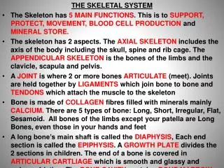
THE SKELETAL SYSTEM • The Skeleton has 5 MAIN FUNCTIONS. This is to SUPPORT, PROTECT, MOVEMENT, BLOOD CELL PRODUCTION and MINERAL STORE. • The skeleton has 2 aspects. The AXIAL SKELETON includes the axis of the body including the skull, spine and rib cage. The APPENDICULAR SKELETON is the bones of the limbs and the clavicle, scapula and pelvis. • A JOINT is where 2 or more bones ARTICULATE (meet). Joints are held together by LIGAMENTS which join bone to bone and TENDONS which attach the muscle to the skeleton • Bone is made of COLLAGEN fibres filled with minerals mainly CALCIUM. There are 5 types of bone: Long, Short, Irregular, Flat, Sesamoid. All bones of the limbs except your patella are Long Bones, even those in your hands and feet • A long bone’s main shaft is called the DIAPHYSIS, Each end section is called the EPIPHYSIS. A GROWTH PLATE divides the 2 sections in children. The end of a bone is covered in ARTICULAR CARTLIAGE which is smooth and glassy and reduces friction. The BONE CAVITY contains BONE MARROW where red blood cells are produced.
THE SKELETAL SYSTEM • Label the Skeleton with the main bones • Colour the Axial and Appendicular Skeletons • Classify each bone as either Long, Short, Irregular, Flat or Sesamoid • Label the elements of a typical Long Bone • Complete the worksheet which identifies Articulating Bones at a Joint • Discuss the functions of the skeleton in your chosen sport • Describe how Cartilage becomes bone in children
There are 3 planes of the body. 1) ANTERIOR – towards the front of the body POSTERIOR – towards the back of the body 2) SUPERIOR – towards the head or upper part of the body INFERIOR – towards the feet or lower part of the body 3) MEDIAL – towards the middle of the body LATERAL – towards the outside of the body The ANATOMICAL POSITION is standing upright with palms face forward and by your side Stand in the ANATOMICAL POSITION and move your synovial (freely moveable) joints in ways that represent the planes above. Can you also name parts of the body that are in relation to the planes. EG: The Pectoralis muscle is anterior of the rib cage and superior of the abdominals
JOINTS • Joints are the area where 2 or more bones ARTICULATE. They are CLASSIFIED in 3 ways according to their balance, stability and mobility • There are FIBROUS (fixed) joints, CARTILAGINOUS (slightly moveable) joints and SYNOVIAL (freely moveable) joints • SYNOVIAL joints have distinguishing features • LIGAMENTS – strong fibrous tissue which join bone to bone • SYNOVIAL MEMBRANE – releases synovial fluid • SYNOVIAL FLUID –lubricates the joint to reduce friction in the JOINT CAVITY • ARTICULAR CARTILAGE – glassy smooth cartilage that absorbs shock and reduces friction • JOINT CAPSULE – tough fibrous tissue which encapsulates and strengthens the joint • BURSA/E – sacks of synovial fluid which prevent ligaments and tendons rubbing • MENISCUS – white fibrocartilage which helps the joint fit together making it stable • PAD OF FAT – provides cushioning between the bone and muscle Critically evaluate the functions of the Structural aspects of a Synovial Joint in the Knee when a basketball player lands after a slam dunk: Mobility and Stability! Explain what would happen if he sprained his knee joint on landing
JOINT MOVEMENT • FLEXION is when the angle of a joint decreases • EXTENSION is when the angle of the joint increases • HORIZONTAL FLEXION is when the shoulder moves across the front of the body • HORIZONTAL EXTENSION is when the shoulder moves laterally from the body • ABDUCTION is movement of the hip or shoulder away from the body – lateral • ADDUCTION is movement of the hip or shoulder towards the body – medial • ROTATION is when the hip or shoulder move around its axis from the ball and socket. This can be MEDIAL and LATERAL. • CIRCUMDUCTION is when the limb moves in a cone shape or the end moves in a circular shape • PRONATION is when the Radio Ulnar joint rotates so the palms face down • SUPINATION is when the Radio Ulnar joint rotates so the palms face up (Bowl of Soup.. ination) • LATERAL FLEXION is when the spine bends sideways when you are in the anatomical position • DORSI FLEXION is when the ankle joint flexes so that the toes point towards the face • PLANTAR FLEXION is when the ankle joint flexes so the toes point down
BICEP BRACHII and TRICEP BRACHII • TRAPEZIUS • PECTORALIS MAJOR • PRONATOR TERES – responsible for Pronation in the forearm • WRIST FLEXORS – inferior of PronatorTeres and WRIST EXTENSORS posterior of the flexors • RECTUS ABDOMINUS • EXTERNAL OBLIQUE – Anterior of Internal Oblique and Lateral of Rectus Abdominus • INTERNAL OBLIQUE – Posterior of External Oblique and Lateral of Rectus Abdominus • Deltoids – 3 – ANTERIOR, POSTERIOR and MIDDLE DELTOIDS • ILIOPSAOS – anterior of femur at superior end of femur • ADDUCTOR LONGUS – Medial of Quadriceps on anterior view • Quadriceps – RECTUS FEMORIS VASTUS LATERALIS VASTUS INTERMEDIUS and VASTUS MEDIALIS • GASTROCNEMIUS • SOLEUS – Inferior and lateral of Gastrocnemius • TIBILIAS ANTERIOR – Lateral of Tibia • Hamstrings– SEMIMEBROSUS (Medial) , SEMITENDONOUS, BICEP FEMORIS (Lateral) • ADDUCTOR MAGNUS – Medial and Posterior of Femur and superior end of femur • GLUTEUS MEDIUS which is superior and lateral to the bigger GLUTEUS MAXIMUS • LATTISIMUS DORSI • INFRASPINATUS which is superior and medial of the TERES MAJOR which is superior of the LattisimusDorsi
The Muscular System • Muscles attach to the skeleton through tendons. There are 2 types of tendon. The ORIGIN tendon is the tendon that remains fixed when the muscle contracts. The INSERTION tendon moves when the muscle contracts. • Muscles work as pairs to move or control a joint. EG: The BICEP BRACHII and the TRICEP BRACHII control the ELBOW joint. The muscle responsible for moving the joint is called the PRIME MOVER or the AGONIST. The other muscle is known as the ANTAGONIST. Together they are known as ANTAGONISTIC PAIRS. • The shoulder muscle … the DELTOID has 3 parts. The ANTERIOR, POSTERIOR and MIDDLE. Each has different functions. • The ROTATOR CUFFS consist of 4 muscles which stabilise the shoulder joint. They are the SUPRASPINATUS, INFRASPINATUS, TERES MINOR and SUBSCAPULARIS. They prevent the larger muscles from displacing the head of the humerus during activity.
Antagonistic Muscle Pairs Extension: Complete the final column only after you have finished all of the other columns
MUSCULAR CONTRACTION • Muscles contract when a stimulus is sent to the muscle which produces tension • There are 2 classifications of muscles contraction. It is either ISOTONIC (changes length) or ISOMETRIC (remains same length) • ISOTONIC contraction can be either CONCENTRIC (the muscle shortens) or ECCENTRIC (the muscle lengthens) • ECCENTRIC contraction occurs when we need to CONTROL a movement against gravity or against another resistance. ISOTONIC …. If we concentrate on the elbow joint. Explain muscle contraction during a bicep curl. Complete the table with your partner. 1st row = Upward Phase. 2nd row = Downward Phase.
MUSCULAR CONTRACTION • Now try this with a PRESS UP using the elbow. Use Row 1 for Upward Phases and Row 2 for Downward Phase PRESS UP Can you now do for the SIT UP ….. A Vertebral joint
MUSCLE FIBRES A MUSCLE FIBRE …is a long cylindrical muscle cell held together in BUNDLES to make up an individual muscle There are 2 main types: 1) SLOW TWITCH FIBRES associated with Aerobic athletes 2) FAST TWITCH FIBRES associated with Anaerobic athletes A) Fast OXIDATIVE GLYCOLITIC Fibres (FOG) – O2 supply B) Fast GLYCOLITIC Fibres – NO O2 Supply MITOCHONDRIA – the power houses or Oxygen Factories of muscle cells. Needed to synthesis O2 for energy MYOGLOBIN– stores and transports O2 in the muscle cell PC - PHOSPHO CREATINE….this bond splits in the muscle to allow quick energy but there is only approximately 10 seconds worth of PC in the muscle GLYCOGEN is stored Glucose or Carbohydrate TRIGLYCERIDE is stored Fat
WARM UP : 3 Phases • PULSE RAISER • MOBILITY EXERCISES– controlled joint movements which rehearse movement patterns • STRETCHES Now explain the impact this will have on the Skeletal Muscle . Think about: Muscle temp / O2 Dissociation / Nerve Impulse Conduction and Contraction / Muscle Force, speed and reactions / Elasticity of Muscles / Muscle Viscosity / Flexibility COOL DOWN : 2 Phases • ACTIVE RECOVERY - Pulse Lowering • STRETCHING – active muscles Now explain the benefits of this on the Skeletal Muscle Tissue: Think about Muscle Temperature, Length of Muscles, DOMS risk, Removal of Lactic Acid,
OSTEOPOROSIS is a weakening of bones caused by a reduction in bone density making them prone to fracture. Often associated with old women but not exclusive to this group. RISK FACTORS include inactivity especially in childhood, and sedentary lifestyles. • PHYSICAL ACTIVITY and HEALTHY DIET are the best defence against it. HIGH IMPACT ACTIVITY is effective in gaining PEAK BONE DENSITY • GROWTH PLATE is the delicate area found between the Diaphysis and the Epiphysis in children. It closes in adult hood. Injuries in this area are common because it is weak. They can lead to weaknesses in adulthood. Injuries are caused through Impact or REPETITION exercises. • OSTEOARTHRITIS is the breakdown and eventual loss of Articular Cartilage. This results in additional friction, loss of flexibility and pain. Its a DEGENERATIVE disease and is caused by repetitive use of joints or Impact. Exercise can have a positive benefit to this ailment. • JOINT STABILITY is the resistance offered by the MusculoSkelatal Tissue that surround joints. It depends on the Joint Shape, its Ligaments and the Muscle Tone. Exercise strengthens these structures but high impact can make weak • MUSCLE POSTURE is when you can carry out activity with efficiency. The muscles responsible are in the CORE. Muscle Tone and Body Weight are is very important
WARM UP AND COOL DOWN Devise a warm up and a cool down for an activity of your choice. Follow the models: Warm Up: 3 Phases • Pulse raiser • Mobility – controlled joint movements which rehearse movement patterns • Stretches Now explain the impact this will have on the Cardio Respiratory Systems and the Musculo - Skeletal Systems. Think about: muscle temp / O2 Dissociation / Nerve Impulse Conduction and Contraction / Muscle Force, speed and reactions / Synovial Fluid / Elasticity of Muscles / Distribution of Blood (Vascular Shunt) / Enzyme Activity for cell Respiration Cool Down: 2 Phases • Active Recovery / Pulse Lowering • Stretch Active Muscles Now explain the benefits of this to; Q (Cardiac Output), Venous Return (VR), Stroke Volume (SV), Minute Ventilation (VE), Blood Pressure, Muscle Temperature, Length of Muscles, DOMS risk, Removal of Lactic Acid, Blood Pooling
MOTION • MOTION is divided into 3 main categories • LINEAR MOTION is when a body moves in a straight or curved line with all the aspects moving at the same speed, at the same time and in the same direction YouTube - 2007 Skeleton & Bobsled World Cup YouTube - SHOT PUT MEN - FINAL 2) ANGULAR MOTION is when a body or part of a body moves in a circle or part of a circle about a point called the AXIS OF ROTATION YouTube - NastiaLiukin 2008 Beijing Olympics Team Finals- Uneven Bars BBC SPORT | Other sport... | Cycling | World record for women pursuit team 3) GENERAL MOTION is a combination of LINEAR and ANGULAR MOTION YouTube - Contraversial Wheelchair Crash YouTube - ATHLETICS -- THROWS JAVELIN WOMEN – FINAL Identify different types of motion in your sport, and then rugby, golf, cricket, track and field
FORCE ….Can perform the following functions • Cause a RESTING body to MOVE • Cause a MOVING body to change DIRECTION, ACCELERATE or DECELERATE • Change an objects SHAPE NEWTONS LAWS OF MOTION 1ST LAW OF MOTION ….A body continues in a state of rest or uniform velocity (same speed and direction) unless acted upon by an external force 2ND LAW OF MOTION – THE LAW OF ACCELERATION: When a force acts upon an object, the rate of change of momentum experienced by the object is proportional to the size of the force and takes place in the direction in which the force acts 3RD LAW OF MOTION….For every action, there is an equal and opposite reaction
CENTRE OF MASS Mass ….is the amount of material of which a body is made. A football is bigger than a shot putt but the shot putt has a greater mass, because the material is heavier. Centre of Mass…..the point where all of the mass is concentrated. The point in which it is balanced in all directions Pin point where you think the Centre of Mass is in the following objects or bodies
STABILITY ….How stable a body or object is! How difficult it is to disturb a body from its balanced position Stability depends on 3 Mechanical Principles 1) Position of the Athletes Centre of Mass 2) Position of the Athletes Line of Gravity 3) Size of the Athlete’s Area of Support
LINE OF GRAVITY Line of Gravity ….is the point from the centre of mass vertically down to the ground
STABILITY CONTINUUM Unstable Less Stable Stable Plot on the line as many different sporting techniques as you can. Make sure you can justify them using the following principles 1) Position of the Athletes Centre of Mass 2) Position of the Athletes Line of Gravity 3) Size of the Athlete’s Area of Support
RELATIONSHIP BETWEEN CENTRE OF MASS AND APPLICATION OF FORCE When a force is applied, the direction of this force in relation to an objects centre of mass will determine whether the motion will be angular motion or linear motion Direct Force…when the force applied goes through the centre of mass Eccentric Force…when the force applied passes outside of the centre of mass causing the resulting motion to be angular Watch the clip and explain this using a Free Kick http://www.youtube.com/watch?v=Pb2qykj6_ZU
CONDUCTION SYSTEM • This is the ELECTRICAL IMPULSE responsible for stimulating the heart to contract. The heart is MYOGENIC (stimulates its own impulse) . 1) The impulse originates in the SINO ATRIAL (SA) NODE located in the posterior wall of the right atrium. It is the PACEMAKER and travels through the left and right atrial walls • Both ATRIA then CONTRACT. The Ventricles do not at this point because they are INSULATED . The impulse makes it way to the ATRIO VENTRICULAR (AV) NODE in the inferior aspect of the right atrium • When the atria finish contracting the Impulse is then sent down the BUNDLE OF HIS fibres which run down the Septum and into the PURKINJE FIBRES which surround the left and right ventricle. When the impulse reaches these both VENTRICLES CONTRACT • Both Ventricles RELAX and the next IMPULSE is released from the SA Node
THE CARDIAC CYCLE • This is the events of ONE HEART BEAT. At rest it lasts 0.8 seconds and is repeated approximately x 72 times per minute. There are 2 Phases A) DIASTOLE is The RELAXATION phase lasting 0.5 seconds B) SYSTOLE is the CONTRACTION phase lasting 0.3 seconds. There are 2 parts to Systole. ATRIAL SYSTOLE and VENTRICULAR SYSTOLE DIASTOLE (0.5 seconds) • Both Atria fill with blood. AV valves are closed • Atrial Blood Pressure rises above Ventricular Pressure • Increased blood pressure forces AV valves open. Blood eases into Ventricles SYSTOLE (0.3 seconds) • Both atria contract FORCING the remaining atrial blood into ventricles • Semi Lunar Valves Remain Closed Both of these are ATRIAL SYSTOLE • Both Ventricles contract increasing ventricular pressure • Aortic and Pulmonary Valves forced open. AV valves closed • Blood is forced into the Aorta and the Pulmonary Artery • Diastole of next cycle begins. SL Valves close stopping backflow of blood THESE 4 ASPECTS MAKE UP VENTRICULAR SYSTOLE
RESTING HEART VALUES • If we measure the hearts OUTPUT we can measure its PERFORMANCE. • HEART RATE is the amount of times the VENTRICLES beat in 1 minute. The average is approximately 72. BRADYCARDIA is when the rate is below 60. This is because of a large STROKE VOLUME because there has been HYPERTROPHY of the VENTRICULAR WALL. • STROKE VOLUME is the amount of blood ejected when a ventricle CONTRACTS. It is the difference of the blood in the ventricle before the contraction (END DIASTOLIC VOLUME) and the volume of blood in the ventricle AFTER the contraction (END SYSTOLIC VOLUME). Therefore STROKE VOLUME = EDV – ESV measured in millilitres • CARDIAC OUTPUT (Q) is the RELATIONSHIP between HR and SV Q (L/min) = STROKE VOLUME (ml per beat) X HEART RATE (BPMs) 1) If your EDV is 130ml and your ESV is 60ml calculate your Stroke Volume. If your Heart Rate is 72 BPMs use this to calculate your Q. 2) During the season your Q is 5L/min but your Resting Heart Rate is 60 BPM, what is your resting SV? Explain why your SV may have increased. 3) Explain why HR decreases and SV increases in Trained athletes?
CARDIO VASCULAR VALUES DURING EXERCISE- STROKE VOLUME • During exercise SV increases LINEARLY until the athlete reaches 40-60% of their maximum speed. Then it PLATEAUS which means that MAXIMAL Stroke Volume values are reached during SUB MAXIMAL (Aerobic) exercise. Stroke Volume increases from approximately 70ml to 120-140ml. • Stroke Volume can INCREASE but it depends on 2 factors • VENOUS RETURN (blood returning to heart). VR reduces in High Intensity • The VENTRICLE’S ability to STRETCH • The heart’s capacity to EMPTY depends on 2 factors • Increased EDV stretches the walls • This increased stretch increases the VENTRICULAR CONTRACTILITY forcing the blood out of the Ventricles when only 50% is pumped out at rest A) An athlete who starts to work towards their MAXIMAL LEVEL (Aerobic Threshold). They have reached their maximal SV. How can they possibly get their Q to increase and continue exercising? Explain also why the athlete may see a reduction in SV as he reaches his maximal intensity B) Explain why SV is reached during sub maximal exercise, and why it even decreases just before maximal intensity is reached C) Draw a graph to represent SV increase. X axis = speed Y Axis = millilitres
CARDIO VASCULAR VALUES DURING EXERCISE- HEART RATE • Heart rate increases just before exercise. This is called the ANTICIPATORY RISE and is caused by a rush of ADRENALIN which stimulates the SA node • Heart Rate increases and decreases as EXERCISE INTENSITY increases and decreases • Heart Rate SLOWS down just before MAX Heart Rate is reached • Heart Rate reaches a PLATEAU during SUB MAXIMAL (Aerobic) exercise. This is called a STEADY STATE. • Heart Rate DECREASES RAPIDLY after exercise during RECOVERY because of reduced demand for O2 • During recovery it will stay above Resting Values to allow the body to repay its OXYGEN DEBT. The greater the O2 Debt, the longer the recovery Draw a graph to represent 2 performers. One is an athlete who has gone jogging at medium intensity for 30 minutes over even terrain. The second athlete is training for 30 minutes over increasingly difficult terrain at medium to high intensity. They are both male 16 years olds. The Y axis should represent Heart Rate from 0 to 200 BPMs and the X Axis represents 3 aspects of time 1) Prior to exercise 0 to 10 minutes 2) Exercise 30 minutes 3) Recovery 30 minutes Label all the above concepts on your graph
CARDIO VASCULAR VALUES DURING EXERCISE- CARDIAC OUTPUT • Q increases in a LINEAR relationship with exercise intensity from RESTING VALUES of approximately 5L/min to MAXIMAL VALUES of between 20 to 40L/min in Highly Trained Endurance athletes • When an athlete reaches 40 – 60% of his MAXIMAL INTENSITY his Stroke Volume PLATEAUS. Therefore any further increases in Q are because of INCREASED HEART RATE • The CARDIOVASCULAR DRIFT is the gradual decrease in SV and increase in HR during prolonged exercise 1) Explain what causes the Cardio Vascular Drift 2) Use the Units given to draw 3 graphs of the response to exercise of Cardiac Output, Stroke Volume and Heart Rate. The X Axis represents Time – Before Exercise, Exercise and Recovery. The Y Axis should represent the units each of the above is measured in
THE CARDIAC CONTROL CENTRE (CCC) • The heart is CONTROLLED and REGULATED by the CARDIAC CONTROL CENTRE situated in the MEDULLA OBLONGATA in the brain • It is controlled by the AUTONOMIC NERVOUS SYSTEM (ANS) which is INVOLUNTARY and consists of SENSORY NERVES (these transmit information from sense receptors to the CCC) or MOTOR NERVES (these transmit information from the muscles to the CCC ) • Sensory Nerves and Motor Nerves can both be part of the SYMPATHETIC NERVOUS SYSTEM (this INCREASES heart rate using the ACCELERATOR NERVE) or PARASYMPATHETIC NERVOUS SYSTEM (this DECREASES heart rate using the VAGUS NERVE). They can do this to your heart rate by sending information to the SA NODE to increase or decrease its IMPULSE. • The CCC receives information about 3 FACTORS: 1) NEURAL CONTROL: information sent by PROPRIOCEPTORS (in joints, muscles, and tendons which detect MOVEMENT), and by CHEMORECEPTORS (which detect pH and 02 & C02 changes in the muscles and aorta) and BAROCEPTORS (which sends info about blood vessel width) 2) HORMONAL CONTROL: This detects ADRENALIN levels in the body 3) INTRINSIC CONTROL: This detects TEMPERATURE and VENOUS RETURN. Both can increase or decrease stimulation of the SA Node
VENOUS RETURN • Blood vessels have 3 layers except capillaries .They are 1 cell thick for 02/C02 DIFFUSION and GASEOUS EXCHANGE. • ARTERIES and ARTERIOLES can VASODILATE (widen) or VASOCONSTRICT (narrow) to allow more or to restrict blood flow • ARTERIOLES have PRE CAPILLARY SPHINCTERS which can close or open to allow blood flow into the capillaries or to shut it off • Large VEINS have VALVES to prevent the backflow of blood. Veins and Venules can VENOCONSTRICT (narrow) and VENODILATE (widen) • VENOUS RETURN is the transport of blood from the capillaries through VENULES into VEINS and back to the heart’s RIGHT ATRIUM • STARLINGS LAW states that Stroke Volume depends on the volume of VR. There are 5 VENOUS RETURN MECHANISMS which help maintain VR • POCKET VALVES: Prevent the backflow of blood • MUSCLE PUMP: Veins are situated near muscle. When the muscle contracts it squeezes the vein and moves the blood along • RESPIRATORY PUMP: The same thing happens in the Chest Cavity • SMOOTH MUSCLE: This is the contraction of the muscles inside the veins • GRAVITY: Blood in the Upper Body returns to the heart more easily
VASCULAR SHUNT MECHANISM • At REST Q (Cardiac Output) is distributed as follows: 15 – 20% is supplied to the MUSCLES and 80 – 85% supplies the ORGANS • During EXERCISE Q is REDISTRIBUTED: 80 – 85% to the WORKING MUSCLES, Organs have reduced supply except the BRAIN. In MODERATE EXERCISE blood flow to the SKIN increases (to reduce TEMPERATURE) but in HIGH INTENSITY it REDUCES (because the need for O2 is greater) • The DISTRIBUTION and REDISTRIBUTION of blood is controlled by the VASOMOTOR CONTROL CENTRE (VCC) It works like the CCC. It is also in the medulla oblongata. It receives information from BAROCEPTORS and CHEMORECEPTORS and the VCC then VASO CONSTRICTS or VASODILATES the ARTERIOLES & PRE CAPILLARY SPHINCTERS • Blood vessels have 3 layers except capillaries .They are 1 cell thick for 02/C02 DIFFUSION and GASEOUS EXCHANGE. • ARTERIES and ARTERIOLES can VASODILATE (widen) or VASOCONSTRICT (narrow) to allow more or to restrict blood flow • ARTERIOLES have PRE CAPILLARY SPHINCTERS which can close or open to allow blood flow into the capillaries or to shut it off • Large VEINS have VALVES to prevent the backflow of blood. Veins and Venules can VENOCONSTRICT (narrow) and VENODILATE (widen)
02 AND C02 TRANSPORT • Blood carries 02 and C02. Oxygen is transported in two ways: • 97% is transported in RED BLOOD CELLS on HEAMOGLOBIN(Hb). It has a HIGH AFFINITY for 02. Each molecule one can carry 4 molecules of 02. This is called OXYHEAMOGLOBIN ( Hb02) 2) 3% is carried in the PLASMA • C02 is transported in 3 ways: • 70% combines with water in red blood cells as CARBONIC ACID • 23% carried on HEAMOGLOBIN as CARBAMINOHEAMOGLOBIN (HbC02) • 7% is dissolved in the PLASMA • Efficient 02 and C02 transport can prolong AEROBIC and ANAEROBIC exercise, delay the ANAEROBIC THRESHOLD, and speed up RECOVERY • A WARM UP speeds up the VASCULAR SHUNT MECHANISM, increase body TEMPERATURE (which increases ENZYME production for muscle contraction and decreases BLOOD VISCOSITY improving blood flow) It delays OBLA (The Onset of Blood Lactate Accumulation – this is point at which the body produces LACTIC ACID faster than it can get remove it) • COOL DOWN prevents POOLING, helps VR, SV,Q and removes LACTIC ACID • Cigarettes contains CARBON MONOXIDE (CO). Hb has a higher AFFINITY to CO than 02 by over 240 times. VEHICLE POLLUTION also contains CO.
BLOOD PRESSURE (Bp) • Blood Pressure is the PRESSURE exerted by the BLOOD against the BLOOD VESSEL walls. It is usually calculated as: = SYSTOLIC Bp / DIASTOLIC Bp • The average pressure is 120mmHg / 80 mmHg (millilitres of mercury) in the Aorta. It can also be expressed as: = BLOOD FLOW (Q) X RESISTANCE. The resistance is the FRICTION of the blood cells against the vessel wall. This is called VISCOSITY (fluid friction) • VASODILATION and VASOCONSTRICTION can decrease and increase Bp. It is measured using a SPHYGMOMANOMETER • Factors such as STRESS, DIET can affect Bp. EXERCISE can also affect it • In ENDURANCE training Systolic Pressure increases LINEARLY to the intensity. Diastolic pressure changes little during SUB MAX exercise but may DECREASE in LOCALISED areas EG: In specific working muscles • In RESISTANCE training powerful muscle contractions BLOCK blood flow. This INCREASES exercise systolic and diastolic Bp, but not resting Bp • In RECOVERY systolic and diastolic Bp REDUCES below resting Bp temporarily. This can have an effect on reducing HYPERTENSION • HYPERTENSION is prolonged high blood pressure which causes CORONARY HEART DISEASES(CHD). It is associated with SEDENTARY LIFESTYLES
IMPACT OF PHYSICAL ACTIVITY ON THE CV SYSTEM • CORONARY HEART DISEASES – 4 (CHDs) are the single largest cause of death in the western world. It is 3 times more likely in SEDENTARY people. • ARTERIOSCLEROSIS is the loss of ELASTICITY, and HARDENING of the blood vessel. This reduces the ability of the vessel to VASODILATE and VASOCONSTRICT . It affects your Bp and VASCULAR SHUNT. Smoking can accelerate this process • ATHEROSCLEROSIS is a form of the above which lines the arteries with CHOLESTEROL and FATTY DEPOSITS causing PLAQUE. This reduces the width (LUMEN) of the vessel and increases risk of BLOOD CLOTS as it causes a high Bp, HYPERTENSION and decreases BLOOD FLOW • HEART: ANGINA is a partial blockage of the CORONARY ARTERY reducing 02 supply to the VENTRICULAR and ATRIAL walls • HEART ATTACK: is a severe, sudden or total RESTRICTION of 02 to the Heart Muscle Wall. It can cause permanent damage and death. It is usually a result of BLOOD CLOTS. • PHYSICAL ACTIVITY protects us from this risk. IMPROVED DIET, refraining from SMOKING. If you have 1 risk factor you may double your chance of CHD, but 3 risk factors increase the risk by 5 times • The WORLD HEALTH ORGANISATION makes many recommendations
IMPACT OF PHYSICAL ACTIVITY ON THE CV SYSTEM We can LESSEN the RISK of CHD. • Improving Heart HYPERTROPHY and VASCULARISATION (increase size and capacity of coronary circulation) • Decreasing BLOOD FIBRINOGEN which reduces clotting • Decreasing BLOOD LIPIDS (triglyceride and cholesterol) • Decreasing LOW DENSITY LIPOPROTEINS (LDL) – high in cholesterol • Increase HIGH DENSITY LIPOPROTEINS (HDL) – these scavenge the cholesterol in the blood • Lower resting Bp and reduce HYPERTENSION • Reduce OBESITY • Alleviate STRESS
MECHANICS OF RESPIRATION • The 3 main FUNCTIONS of the Respiratory System are 1) PULMONARY VENTILATION (breathing air in and out) 2) EXTERNAL RESPIRATION is the exchange of 02 and C02 at the ALVEOLI 3) INTERNAL RESPIRATION is the exchange of 02 and C02 between the MUSCLES and BLOOD • INSPIRATION is breathing in and EXPIRATION is breathing out. Changes in lung VOLUME is initiated by the RESPIRATORY MUSCLES. 5 STEPS: • Muscles ACTIVELY contract or PASSIVELY relax which cause .. • MOVEMENT of the ribs, sternum and abdomen which cause... • The THORACIC CAVITY to increase or decrease in VOLUME which causes . • The LUNG AIR PRESSURE to increase or decrease which causes .. • INSPIRATION or EXPIRATION to occur • At REST the muscles that CONTRACT to cause INSPIRATION are the DIAPHRAGM and the EXTERNAL INTERCOSTAL muscles. During EXPIRATION at REST these muscles relax and are PASSIVE • During EXERCISE we have to recruit the STERNOCLEIDOMASTOID, SCALENES and PECTORALIS MINOR in INSPIRATION. In EXPIRATION Ex Intercostals & Diaphragm STILL relax BUT we now CONTRACT the INTERNAL INTERCOSTALS ,RECTUS ABDOMINUS and OBLIQUES
LUNG VOLUMES • TIDAL VOLUME (TV) is the volume of air INSPIRED or EXPIRED per BREATH. At REST this is approximately 500ml. During EXERCISE this can increase by 3 / 4 litres • FREQUENCY (f) is the number of breaths taken in one minute. Sometimes referred to as RESPIRATORY RATE. At REST it is approximately 12 – 15. During EXERCISE this can increase to 40 to 60 breaths • MINUTE VENTILATION (VE) is the VOLUME of air INSPIRED or EXPIRED in one minute. At REST this is approximately 6 to 7.5 L/min. During EXERCISE this can increase to between 120 to 180 L/min if trained • VE = TV x f • During SUB MAXIMAL exercise TV and f increase to allow for a greater VE but during MAXIMAL INTENSITY exercise only an increase in f will allow for more VE because it takes too long in time to increase TV 1) If an athlete has a resting TV value of 500ml and a respiratory frequency of 12, calculate their VE 2) Explain why the athlete should increase their f to 24 and their TV to 4000ml per breath during exercise would be beneficial to an aerobic athlete
GASEOUS EXCHANGE • Gaseous Exchange is the exchange of 02 and C02. It relies on DIFFUSION which is movement of gases from an area of HIGH PRESSURE to an area of LOW PRESSURE. Gases ALWAYS do this. The difference between these 2 pressures is called the DIFFUSION GRADIENT. The BIGGER the gradient, the more DIFFUSION and GASEOUS EXCHANGE can occur. • The PARTIAL PRESSURE (PP) of gases is the amount of pressure a gas exerts on other gases in the same area. EG: When blood is OXYGENTATED it has a HIGH PARTIAL PRESSURE (PP) of 02 and a LOW PARTIAL PRESSURE (PP) of C02. The opposite is true in DEOXYGENATED blood. • PARTIAL PRESURES can occur in 2 locations. • EXTERNAL RESPIRATION: This is at the ALVEOLI. HIGH PP of 02 diffuse from the alveoli to the capillary where the PP of 02 is LOW. Similarly a HIGH PP of C02 in the capillary diffuses to the alveoli where the PP of C02 is LOW • INTERNAL RESPIRATION: This is at the point where the capillaries surround the MUSCLES. A HIGH PP of 02 in the capillaries diffuse into the muscle where there is a LOW PP of 02. Similarly a HIGH PP of C02 in the muscles diffuse out to the capillaries where the PP of C02 is LOW
GASEOUS EXCHANGE DURING EXERCISE • Both Internal and External Respiration INCREASE during exercise to supply the 02 to the working muscles • The OXYGEN – HAEMOGLOBIN DISSOCIATION CURVE is a graph which informs us about how much HAEMOGLOBIN is SATURATED with 02 in both the LUNGS and in the MUSCLE TISSUE. Dissociation is when 02 unloads from Haemoglobin. • At REST: In EXTERNAL respiration there is a PP of 02 of 100mmHg in the alveoli. The blood then travels to the muscles. In INTERNAL respiration at the muscles the PP of 02 is 40mmHg Draw the Graph. The X Axis has Units of mmHg ranging from 0 to 100mmHg. The Y Axis has Units of % 02 Saturation of Haemoglobin ranging from 0 to 100%. Now plot the Curve using the following data: Now label the X Axis to identify PP of 02 in Muscle Tissue = 40mmHg Now label the X Axis to identify PP of 02 in the Lungs = 100 mmHg Now calculate the Percentage Saturation of Haemoglobin in Muscle Tissue and in the Lungs. What is the percentage difference? What has happened?
Therefore at REST approximately 25% of the 02 that is carried by the Haemoglobin is UNLOADED into the muscles. If we can increase this percentage we can have a more efficient athlete who will perform higher • The DIFFUSION GRADIENT is the difference between 2 PPs of the same gas. The bigger the Gradient the more diffusion takes place • During EXERCISE the muscles are using more 02, and are producing more C02. Therefore Deoxygenated blood coming back to the lungs via the heart has a HIGH PP of C02 and a LOW PP of 02. At EXTERNAL RESPIRATION the blood reaches the capillaries that surround the alveoli, this means that C02 diffuses QUICKER into the lungs and out of the body and that 02 diffuses QUICKER into the capillaries because the DIFFUSION GRADIENT is higher. This means that HAEMOGLOBIN is almost FULLY SATURATED • At INTERNAL RESPIRATION 4 factors have the effect of moving the graph line to the right. They are 1) Increase in TEMPERATURE 2) Decrease in PP of 02 in the Muscles 3) Increase in PP of C02 in Muscles 4) BOHR EFFECT which is an increase in ACIDITY because of LACTIC ACID. Plot the graph • Calculate the Percentage uptake now. What has happened?
VENTILATORY RESPONSE TO LIGHT, MODERATE AND HEAVY EXERCISE Draw the following graph: X Axis = Units = Time: 3 parts A) Pre Exercise From -3 minutes to 0 minutes B) Exercise from 0 minutes to 5 minutes C) Recovery from 5 to 7 minutes. Y Axis Units = Minute Volume (VE) from 0 to 140 L/minute. Now plot the following coordinates for the 3 Exercise Lines Annotate the following on your graph: • Identify the ANTICIPATORY RISE • Identify PLATEAUS • Identify SLOW but Continued Increase in VE just before Maximal Intensity • Identify the RAPID DECREASE in VE • Identify a SLOW but gradual DECREASE towards resting VE CAN YOU EXPLAIN HOW THE BODY REGULATES AND CONTROLS THIS
RESPRATORY CONTROL CENTRE (RCC) • The RCC REGULATES and CONTROLS Pulmonary Respiration (Breathing). Like the CCC and the VCC it is also controlled in the medulla oblongata. The Mechanic of Breathing is CONTROLLED by the RESPIRATORY MUSCLES which are INVOLUNTARY muscles. The RCC controls them and has 2 areas • The INSPIRATORY CENTRE This works during both REST AND during EXCERCISE. It sends messages from the RCC along the PHRENIC NERVE to the DIAPHRAGM and along the INTERCOSTAL NERVES to the INTERCOSTAL MUSCLES. When an IMPULSE reaches these muscles they CONTRACT to increase Lung Volume. When they are not stimulated they RELAX which decreases Lung Volume. During Exercise it also stimulates the STERNOCLEIDOMASTOID, the SCALENES and PECTORALIS MINOR • The EXPIRATORY CENTRE only works during EXERCISE. It does this to further INCREASE the DEPTH and FREQUENCY. This centre sends an impulse to the Expiratory Muscles which are the INTERNAL INTERCOSTALS, the RECTUS ABDOMINUS and the OBLIQUES • CHEMORECEPTORS detect increase PP C02, decrease PP 02 and Lactic Acid • PROPRIOCEPTORS detect joint and muscle movement • THERMORECEPTORS detect changes in Temperature • BAROCEPTORS detect stretches in the lungs
ALTITUDE – THE EFFECTS ON THE RESPIRATORY CENTRE • When exercising at ALTITUDE it has a significant effect on respiration. Altitude is height of 1500 ms+ above SEA LEVEL. At altitude the PP of 02 is HYPOXIC. This means it is massively reduced. This decreases performance BUT training at altitude is an ERGOGENIC AID (improves performance) • Due to HYPOXIC conditions the body learns to ADAPT by increasing the bodies amount of EPO which increases your RED BLOOD CELL count. You also gain CAPILLARISATION (an increase in capillaries). The main effect is that when you RETURN to sea level your AEROBIC capacity improves • Altitude Training is EXPENSIVE and research does not support it. Any ADAPTATIONS are SHORT LIVED and are only an advantage for a few days after High Altitudes can actually PREVENT aerobic athletes from training at the same INTENSITY or DURATION and so V02 MAX (Maximum 02 Consumption) is reduced. ANAEROBIC events may benefit however. • There are 3 methods of Altitude Training A) LIVE HIGH TRAIN HIGH (LHTH) B) LIVE LOW TRAIN HIGH (LLTH) C) LIVE HIGH TRAIN LOW (LHTL)
4 ADAPTATIONS TO THE RESPIRATORY SYSTEM • Training increases the bodies ability to transport 02, increase AEROBIC CAPACITY and increase the LACTATE THRESHOLD There are 4 specific ADAPTATIONS • RESPIRATORY STRUCTURES increase number of alveoli, increased elasticity and endurance • BREATHING MECHANICS increase strength, power, endurance and decreased fatigue of the RESPIRATORY MUSCLES • RESPIRATORY VOLUMES: Lung Volumes do not change much but TIDAL VOLUME can during MAXIMAL intensity. Maximal VE increases. FREQUENCY decreases at rest but increases during Maximal Intensity • DIFFUSION remains unchanged at rest but increases during maximal exercise • The main PERFORMANCE BENEFITS are improved AEROBIC PERFORMANCE, increased ENDURANCE, delay of ANAEROBIC THRESHOLD, increased EFFICIENCY and more HEALTHY
EFFECTS OF ASTHMA AND SMOKING ON THE RESPIRATORY SYSTEM • ASTHMA is a narrowing of the airways (BRONCHOCONSTRICTION) in response to a TRIGGER or an ALLERGEN. Its symptoms include WHEEZING, BREATHLESSNESS , MUCUS PRODUCTION and COUGHING • It is measured using a SPIROMETER which measures EXHALED air volume. People are said to have asthma if this volume increases with treatment. The IOC allows 15% improvement for 1 sec after treatment • TRIGGERS include drying and water loss in the airways, and exercise. This is called EIA (EXERCISE INDUCED ASTHMA). This is common in cold weather sports where the air is DRY and when pollutants are use on ice rinks. • ALLERGENS include exhaust fumes, pollen, dust, air pollutants and pollens • MEDICATION include ‘Relievers’ coded BLUE which relax airway muscles. A Daily dose of CORTICOSTEROIDS ‘Preventers’ coded non blue suppress chronic inflammation and improve pre exercise lung function. • NON MEDICAL TREATMENTS include a 10 to 30 min WARM UP which provides a 2hourREFRACTORY period. DIET of reduced salt and fish oils both reduce EIA. Caffeine helps but the IOC limit is 12mg/ml. INSPIRATORY MUSCLE TRAINING (IMT) can strengthen the RESPIRATORY MUSCLES • SMOKING impairs development in TEENAGERS, impairs LUNG FUNCTION and increases likelihood of DISEASES and INFECTIONS


