Download
1 / 79
790 likes | 1.04k Views
Peripheral Nervous System: Efferent Division. Autonomic Nervous System Muscle Physiology. Chapter 7 Goals. After studying this chapter, students should be able to . . . 1. compare the structures and pathways of the autonomic system with those involved in the control of skeletal muscle.

E N D
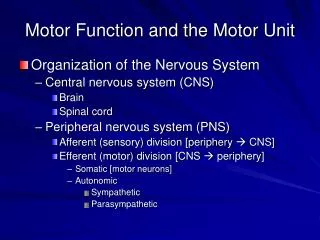
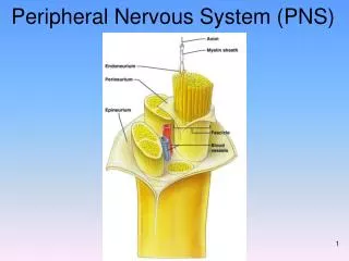
Peripheral Nervous System: Efferent Division • Autonomic Nervous System • Muscle Physiology
Chapter 7 Goals After studying this chapter, students should be able to . . . 1. compare the structures and pathways of the autonomic system with those involved in the control of skeletal muscle. 2. explain how autonomic innervation of involuntary effectors differs from the innervation of skeletal muscle. 3. describe the structure and general functions of the sympathetic division of the autonomic system. 4. describe the structure and general functions of the parasympathetic division of the autonomic system. 5. list the neurotransmitters of the preganglionic and postganglionic neurons of the sympathetic and parasympathetic systems. 6. describe the structural and functional relationships between the sympathetic system and the adrenal medulla. 7. distinguish between the different types of adrenergic receptors and explain the physiological and clinical significance of these receptors.
Chapter 7 Goals After studying this chapter, students should be able to . . . 8. explain how the cholinergic receptors are categorized and describe the effects produced by stimulation of these receptors. 9. explain the antagonistic, complementary, and cooperative effects of sympathetic and parasympathetic innervation on different organs. 10. explain how the autonomic system is controlled by the brain.
Chapter 8 Goals After studying this chapter, students should be able to . . . • describe the gross and microscopic structure of skeletal muscles. • describe the nature of a muscle twitch and explain how summation and tetanus are produced. • distinguish between isometric and isotonic contractions. • explain how the series-elastic components affects muscle contraction. • define the term motor unit and explain how motor units are used to control muscle contraction. • describe the structure of myofibrils and explain how it accounts for the striated appearance of skeletal muscle fibers. • explain what is meant by the sliding filament theory of contraction. • list the events that occur during cross-bridge cycles and describe the role of ATP in muscle contraction. • explain how tropomyosin and troponin control muscle contraction and relaxation, and describe the role of Ca2+ and the sarcoplasmic reticulum in excitation-contraction coupling.
Chapter Goals • describe the structure and function of muscle spindles and explain the mechanisms involved in a stretch reflex. • explain the function of Golgi tendon organs and explain why a slow, gradual muscle stretch could avoid the spasm that may result from a rapid stretch. • explain what is meant by reciprocal innervation and describe the neural pathways involved in a crossed-extensor reflex. • explain the significance of gamma motoneurons in the neural control of muscle contraction and in the maintenance of muscle tone. • explain the significance of the maximal oxygen uptake, and the function of phosphocreatine in muscles. • explain how slow-twitch, fast-twitch, and intermediate fibers differ in structure and function.
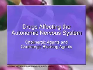
AUTONOMIC NERVOUS SYSTEM • Nerve Pathways • Organization • Receptors • Neuromuscular Junction
ANS RECEPTORS 7-4
IX. MUSCLES • Muscle types • Details of Skeletal Muscle • Mechanism of Contraction • Whole Muscle Physiology
Muscle types • Smooth • Cardiac • Skeletal (=striated)
Muscle Types 8-1
Molecular Composition • Major Proteins • Actin - composed of G-actin subunits " thin filaments • Myosin " thick filaments • Minor Proteins • Troponin • TnT - Tropomyosin-binding subunit • TnC - Calcium-binding subunit • TnI - Inhibitory subunit • Tropomyosin - lies in groove. Spans 7 G-actin subunits
Motor Unit • All the muscle fibers innervated by a single neuron
All-or-none Law • “A muscle fiber contracts to the greatest extent of its immediate capacity or not all all”
Minimal/Maximal Stimulus • Minimal -- the greatest stimulus required to evoke the least response or a stimulus sufficient to recruit a single motor unit • Maximal -- the least stimulus required to evoke the greatest response or a stimulus sufficient to recruit all motor units
Recruitment • When a stimulus exceed the threshold of a motor unit, it will contract (will be recruited) and contribute to the strength of contraction.
Summation • Adding together of the effects of stimuli to cause or increase the magnitude of a response. • a. Temporal - summation in time. Increased frequency of stimulation from a single source. • b. Spatial - summation in space. Two or more sources of stimulation are moved closer together in space.
Tetany • Sustained contraction to a muscle's immediate capacity. Stimuli get closer together, so progressively less time is available for relaxation. Eventually, the muscle will have no time to relax, and will be tetanized
Fatigue • Conduction Failure -- results from K+ build-up in small space of T-tubule • Lactic Acid build-up -- increased H+ changes protein conformation • Inhibition of cross-bridge cycling -- results from build-up of ADP
Treppe • Staircase phenomenon. When a fresh muscle is used, each contraction makes the next contraction more efficient up to a point. Probably a result of increased temperature
Contracture • An upward drifting of a recording's base line as a result of an increased relaxation time as a muscle tires
Neural Control of Skeletal Muscles • Muscle Spindle • Monosynaptic Reflex • Disynaptic Reflex • Reciprocal Innervation • Crossed-extensor Reflex
Spindles • Group I • Annulo-spiral • Phasic (How is stretch changing?) • Group II • Flower spray • Tonic (Where are you before change occurs?)