Download
1 / 15
160 likes | 281 Views
Cellular Control . You should be able to: ( a) state that genes code for polypeptides, including enzymes; (b) explain the meaning of the term genetic code ; (c) describe, with the aid of diagrams, the way in which a nucleotide sequence codes for the amino acid sequence in a polypeptide;

E N D
Cellular Control You should be able to: (a) state that genes code for polypeptides, including enzymes; (b) explain the meaning of the term genetic code; (c) describe, with the aid of diagrams, the way in which a nucleotide sequence codes for the amino acid sequence in a polypeptide; (d) describe, with the aid of diagrams, how the sequence of nucleotides within a gene is used to construct a polypeptide, including the roles of messenger RNA, transfer RNA and ribosomes; (e) state that mutations cause changes to the sequence of nucleotides in DNA molecules; (f) explain how mutations can have beneficial, neutral or harmful effects on the way a protein functions; (g) state that cyclic AMP activates proteins by altering their three-dimensional structure; (h) explain genetic control of protein production in a prokaryote using the lac operon; (i) explain that the genes that control development of body plans are similar in plants, animals and fungi, with reference to homeobox sequences (HSW1); (j) outline how apoptosis (programmed cell death) can act as a mechanism to change body plans.
Key Definitions A gene is a length of DNA that codes for one or more polypeptides. A genome is the entire DNA sequence of that organism. The human genome consists of approximately 3 million nucleotide base pairs. A polypeptide is a polymer consisting of amino acids joined by peptide bonds. A protein is a large polypeptide, usually 100+ amino acids. Some proteins have more than 1 polypeptide chain. Transcription: the first stage in protein synthesis that occurs in the nucleus. It is the creation of a single stranded mRNA copy of the DNA coding strand. Translation: the second stage of protein synthesis which involves the assembly of polypeptides at ribosomes in the cytoplasm. Amino acids are placed in the correct order according to the sequence of codons on the mRNA. Mutations are structural changes to the genetic material within a cell – either to a gene or to a chromosome. Lac Operon: a length of DNA containing a series of genes coding for enzymes and proteins that allow bacteria to use lactose, plus genes regulating their transcription and translation. Regulator Gene: DNA sequence that codes for the Lac Repressor protein Promoter: section of DNA to which the enzyme RNA polymerase binds to begin transcription of structural genes. Operator: section of DNA to which the Lac Repressor protein can bind when lactose is absent B-Galactosidase: codes for an enzyme that breaks down Lactose Lactose Permease: codes for a carrier protein that increases the uptake of lactose into the cell through the plasma membrane. Homeobox genes: control the development of the body plan of an organism, including the polarity and positioning of the organs. Apoptosis: programmed cell death in multicellular organisms.
The sequence of nucleotide bases on a gene provide a code, with instructions for the construction of a polypeptide. This genetic code: • is a triplet code: a sequence of 3 nucleotide bases codes for an amino acid. • is a degenerate code: there is more than 1 triplet code for a particular amino acid. • is widespread: base sequences can code for the same amino acid in all organisms. Genes code for polypeptides. This includes enzymes like rubisco, hormones like adrenaline and insulin and structural proteins like collagen and keratin. It also includes antibodies, haemoglobin, channel proteins, electron carriers and the tubulin proteins of the cytoskeleton. Transcription Translation In order to make a polypeptide, a copy of the genetic code that can pass through a pore in the nuclear envelope into the cytoplasm must be made. This happens through transcription. Transcription creates a single stranded mRNA copy of the DNA coding strand. This mRNA strand must then attach to a ribosome where protein synthesis will occur – this is translation.
1. Transcription • Takes place in the nucleus • One gene is transcribed from DNA to mRNA • Catalysed by enzyme RNA Polymerase Portion of DNA containing the gene to be transcribed dips into the nucleolus and the double helix unwinds at this point only. mRNA nucleotides are activated by having an extra 2 phosphate groups added. mRNA nucleotides align next to the DNA template or ‘sense’ strand according to complementary base pairing. The extra 2 phosphate groups are hydrolysed to release energy used to join the nucleotides to form a strand of mRNA. 4. mRNA leaves the nucleus through a nuclear pore and the DNA double helix rewinds. Hydrolysed Complementary base pairing between DNA and mRNA:
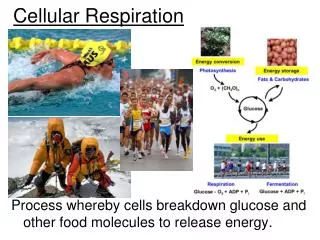
2. Translation • Takes place in the cytoplasm • Assembly of polypeptides at ribosomes • Amino acids are placed in sequence according to the sequence of codons on the mRNA. 5. mRNA molecule attaches to a ribosome in the cytoplasm 6. The ribosome can read 2 mRNA codons at a time 7. tRNA molecules with mRNA codons according to complementary base pairing. tRNA anticodons are complementary to mRNA codons. 8. As tRNA molecules align, this brings amino acids into the correct sequence to form the protein primary structure. 9. Peptide bonds form between the amino acids. tRNA molecules are released and reactivated. 10. Polypeptide chain is activated by cAMP which causes the folding and coiling of the polypeptide chain into a specific shape according to the protein formed.
Why is the amino acid sequence so important? Transfer RNA These 3 unpaired bases are the amino acid binding site. The tRNA molecule will be activated when an amino acid binds here. The amino acid that will bind is determined by the anticodon code. The sequence of amino acids determines the primary structure of the protein. The primary structure determines the tertiary structure – how the protein folds up into its three dimensional shape. tRNA is made in the cytoplasm and passes into the cytoplasm. The RNA folds into hairpin shapes. The tertiary structure is what allows the protein to function. If the tertiary structure is altered, the protein can no longer function. For example, the active site of an enzyme may have an altered shape and the substrate molecules will no longer fit. The anticodon is 3 bases which binds to the complementary codon on the mRNA strand.
Mutations Chromosome mutations A large section of DNA is lost from a chromosome. Sometimes the lost section of DNA reattaches to another chromosome. Whole chromosomes can be duplicated or deleted – an example of this is trisomy 21 which causes Down’s Syndrome. Mutations are important because they mean a change in the nucleotide base sequence. This base sequence determines the amino acid sequence, which in turn determines the tertiary structure and so the function of the protein. If the protein structure is incorrect, it will be non functioning. DNA Mutations This is a change to the sequence of nucleotides along the DNA. DNA mutations can be point/substitution which is when one base is replaced with another or insertion and deletion mutations where a base is added or removed from the genetic code.
Types of DNA Mutation Silent Mutations: The genetic code is degenerate – there is more than one triplet code for each amino acid. Some point mutations will therefore have no effect on the protein produced because the mutated triplet code still codes for the same amino acid. Missense Mutations: Changing one base changes one of the amino acids in the primary structure. Effect on the protein will depend on how important the amino acid is in its structure and how similar the amino acids are to each other. Nonsense Mutations: Changing a base has created a stop codon in the middle of the mRNA. This means that a much shorter and probably non functioning protein will be produced in translation. Frameshift Mutations: Addition or deletion of one base means that the correct reading frame is lost. Many of the amino acids included in the polypeptide are likely to be different, so the protein will probably be non-functioning.
What are the outcomes of mutations? • Some mutations have a neutral effect: they have no apparent advantage or disadvantage to the organism. If a gene is altered by a change to its base sequence, it becomes another version of the same gene: it is an allele of the gene. Examples of this include: • Attached ear lobes/free ear lobes • Ability to roll the tongue Free Ear Lobe Attached Ear Lobe Early humans in Africa had dark skin. Melanin pigment protected them form the harmful effects of UV light. Any humans that had a mutation causing paler skin would have burned and suffered with skin cancer. Early humans with mutations producing paler skin due to a lack of pigment would have an advantage over those with dark skin as humans moved to more temperate climates. Sunlight there was not intense enough to cause vitamin D production in those with dark skin, so they would suffer form rickets. Depending on the environment, the same mutation for paler skin can be beneficial or harmful. When an environment changes, individuals within a population who have a certain characteristic may be better adapted to the new environment. Well adapted organisms can out-compete those who do not have the advantageous characteristic. This is natural selection – the mechanism for evolution.
The Lac Operon Bacteria normally use glucose as their respiratory substrate because it is quick and easy to respire in glycolysis. However, if placed in an environment with no glucose and instead with lactose present, the bacteria will start to respire lactose. Structural Genes Control Sites Regulator Gene Y I Z A P O DNA sequence that codes for the Lac Repressor Protein O is the operator site: this is the section of DNA to which the Lac repressor protein will bind when lactose is absent. P is the promoter site: this is the section of DNA to which RNA Polymerase enzyme binds to begin transcription. Z and Y are structural genes that code for the enzymes B-Galactosidase and Lactose Permease.
Lac operon: Glucose Present; No Lactose 3. RNA Polymerase cannot bind to the promoter region, so the structural genes for B-Galactosidase and Lactose Permeasecannot be transcribed. Y I Z A P O 4. The enzymes cannot be synthesised, so lactose cannot be respired. 2. Lac repressor protein binds to operator region 1. Regulator gene is transcribed and translated and the Lac Repressor Protein is synthesised. The Lac Operon provides a survival advantage to the bacteria. When glucose is present, the E. Coli should use it exclusively, as it is much easier to respire than lactose. No amino acids or ATP is wasted on the production of unnecessary enzymes. When glucose is absent, but lactose is available, the Lac Operon enables the bacterium to continue to respire to release energy.
Lac operon: Glucose absent; Lactose present 3. RNA Polymerase can now bind to the promoter region, so the structural genes for B-Galactosidase and Lactose Permeasecan be transcribed. 1. Regulator gene is transcribed and translated and the Lac Repressor Protein is synthesised. Y I Z A P O 2. Lactose binds to the Lac Repressor protein’s allosteric site. Lactose is a non competitive inhibitor and changes the shape of the repressor protein so that it cannot bind to the operator region. 4. Transcription and translation of structural genes into lac enzymes, allowing the bacterium to respire lactose.
Homeobox Genes All animals possess the same 8 Hox genes: they lie along the chromosome in the same order as they are expressed along the body. Each hox gene is linked to its own set of genes responsible for controlling 1 section of the body. One hox gene can activate and turn on 1000s of other genes. Hox Genes code for proteins called Transcription Factors. These proteins are able to interact with DNA to cause particular genes to be transcribed into mRNA. How do Hox genes work? In all of the cells that will become a head, the ‘head’ hox gene will be switched on. The ‘head’ hox gene is transcribed and translated to make a transcription factor. The transcription factor activates all of the genes in the genome needed to make a head. The ‘head’ genes are transcribed and translated to form head making proteins and enzymes. Duplication of hox genes drives the evolution of complexity. All animals have the same set of basic Hox genes, but body plans will become more complex when hox genes are duplicated. For example, whereas a sponge will have just 1 copy of each hox gene, a mouse will have at least 4. Each duplicate of the gene is free to take a slightly different role to add complexity to the organism’s body plan.
Apoptosis • 1. Enzymesbreak down the cell cytoskeleton. • 2. The plasma membrane changes and small swellings called blebs form. • 3. Chromatin condenses, DNA fragments and the nuclear envelope breaks up. • 4. The cell breaks into vesicles. • 5. Vesicles are taken up by phagocytes, cellular debris is digested and disposed of so it doesn’t damage other cells. • No harmful hydrolytic enzymes are released unlike with necrosis.
Apoptosis and the Hayflick Limit: Normal body cells divide a limited number of times. This is around 50 mitotic divisions. After this, a series of biochemical events leads to orderly cell death. • Apoptosis is vitally important: • In developing a complex body structure: apoptosis allows digits on hands and feet to separate from each other • In removing damaged, infected or unneeded self cells. This is crucial in reducing the risk of cells becoming cancerous. Apoptosis also removed ineffective T Lymphocytes from the immune system which reduced the risk of autoimmune diseases developing. Apoptosis is controlled though cell signals – either an external signal or from within the cell itself. Nitric oxide will make the inner mitochondria membranemore permeable to H+ ions, so the H+ gradient is lost. In response to this, proteins are released which bind to apoptosis inhibitor proteins in the cytoplasm, allowing apoptosis to take place. Too much apoptosis = cell loss and degradation of organs Too little apoptosis = formation of tumours