Download
1 / 111
1.11k likes | 1.14k Views
Discussing present treatment options and future strategies for patients with colorectal cancer while presenting real case studies.

E N D
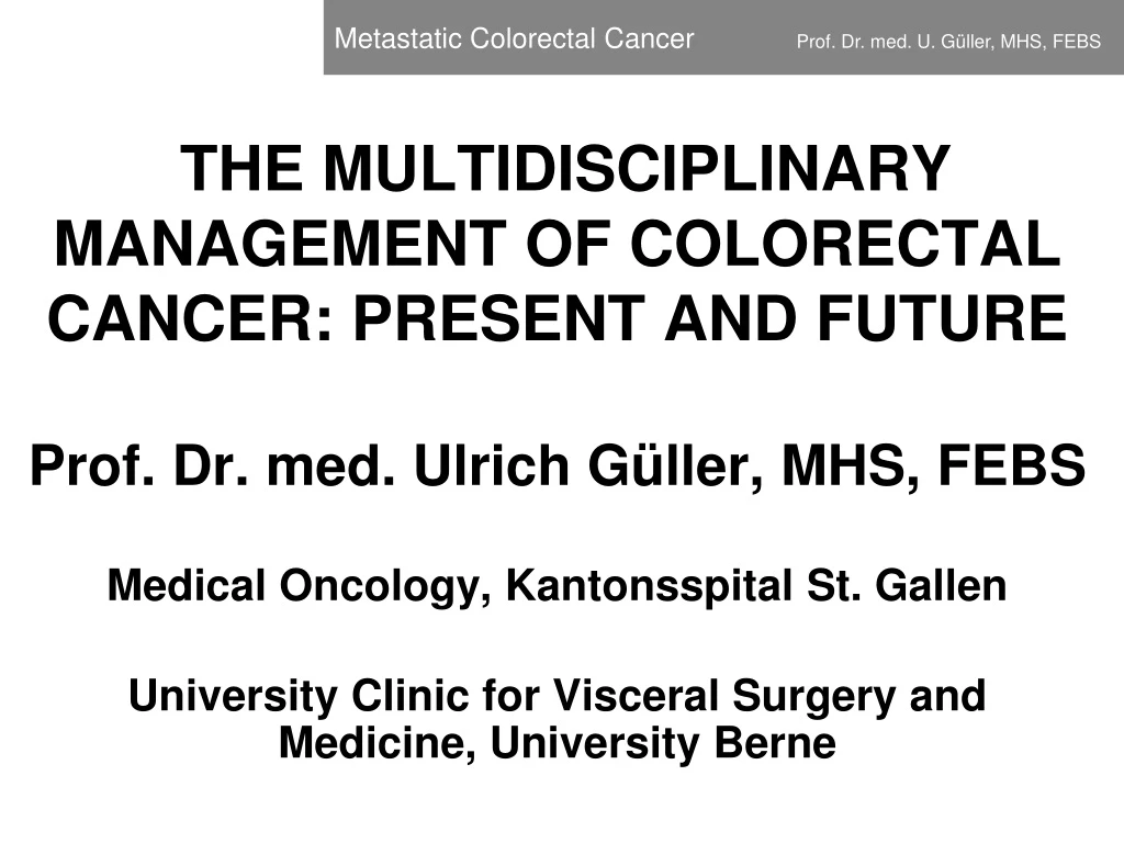
Prof. Dr. med. Ulrich Güller, MHS, FEBS Medical Oncology, Kantonsspital St. Gallen University Clinic for Visceral Surgery and Medicine, University Berne The multidisciplinary management of colorectal cancer: present and future
Overview 3 case presentations Current treatment armamentarium in patients with metastatic colorectal cancer Outlook: future treatment strategies Discussion/Questions
Patient 1 58 yo Male IT Specialist • 58 yo male IT-specialist • No relevant past medical history • Unspecific abdominal pain, fatigue
58 yo Male IT Specialist • Primary care physician: Ultrasound • „Big“ lesion right liver lobe • Computertomographie
Computertomographie • 20cm lesion in right liver lobe • Nootherlesions on CT scan • Thikening of sigmoidcolon
Computertomographie • 20cm lesion in right liver lobe • Nootherlesions on CT scan • Thikening of sigmoidcolon => Colonoscopy + biopsy: Adenocarcinoma G2 sigmoidcolon
Computertomographie • 20cm lesion in right liver lobe • Nootherlesions on CT scan • Thikening of sigmoidcolon • Colonoscopy + biopsy: Adenocarcinoma G2 sigmoidcolon • Sigmoidcarcinomawithsynchronoussingularlivermetastasis
Gastroenterology • Medical Oncology • Radiation-Oncology • Surgical Oncology • Radiology • Interventionel Radiol. (TAE, RFA, MWA, IRE) • Patholology • Nuclear Medicine (SIRT) Multidisciplinary Tumorboard
Gastroenterology • Medical Oncology • Radiation-Oncology • Surgical Oncology • Radiology • Interventionel Radiol. (TAE, RFA, MWA, IRE) • Patholology • Nuclear Medicine (SIRT) • => BRAIN POWER!!! Multidisciplinary Tumorboard
Systemic disease • Synchronous singule liver metastasis from sigmoid cancer Multidisciplinary Tumorboard
Step 1: Inductionchemotherapy Multidisciplinary Tumorboard
Step 1: Inductionchemotherapy Step 2: Resectionliver (liver-first) Multidisciplinary Tumorboard
Step 1: Inductionchemotherapy Step 2: Resectionliver (liver-first) Step 3: Chemotherapy Multidisciplinary Tumorboard
Step 1: Inductionchemotherapy Step 2: Resectionliver (liver-first) Step 3: Chemotherapy Step 4: Oncologicsigmoid resection Multidisciplinary Tumorboard
OCFL- regime • 3 chemotherapeutic agents (5FU, Oxaliplatin, Irinotecan) • Weekly administration • Port-à-Cath • 3 months of treatment Chemotherapy Roth A et al, Ann Oncol 2005
After 3 Months of Chemotherapy Nach Chemotherapie
Prior After Nach Chemo Vor Chemo
Good partial remission • General conditions improved during chemotx • Relevant decrease of tumormarkers • Ca 19-9: 14`200 => 64 • CEA 3`940 => 16 Chemotherapy
Decision: Right-sidedhemi-hepatectomy MD Tumorboard
Liver Specimen Histology: < 10% vital cancercells!
Chemotherapy: 6 x FOLFOX (5FU und Oxaliplatin) • Oncologicsigmoidcancer resection • ypT3, ypN1a (1/17) ypM1a (HEP) • Patient remains in completeremission 5 years after surgery 58-yo Male IT-Specialist
Excellentcollaborationbetween surgicalandmedicaloncologists! 58-yo Male IT-Specialist
Patient 2 55 yo Female Patient
Fatigue, irondeficiency • Colonoscopy • Cancer ascendingcolon • CT: 6 bilobarhepaticmetastases • 1 lesionseg. II, all otherlesions: right lobe • = > Synchronoushepaticmetastases 55 yo Female Patient
Step 1: Inductionchemotherapy Step 2: Clearing liver (R-lobe plus RFA II) Step 3: Chemotherapy Step 4: Righthemicolectomy MD Tumorboard
FOLFOX 6 infusions (Nordlinger/EORTC) • Very good partial remission 55 yo Female Patient
Prior Post
Prior Post
Tumorboard: Neoadjuvant systemic treatment • FOLFOX 6 infusions • Very good partial remission • 5/2015: Tumorboard: Right hemihepatectomy/RFA metastasis seg. II 55 yo Female Patient
Tumorboard: Neoadjuvant systemic treatment • FOLFOX 6 infusions • Very good partial remission • 5/2015: Tumorboard: Right hemihepatectomy/RFA seg. II • Histology: complete path. remission 55 yo Female Patient
Histology: complete pathologic remission • FOLFOX x 6 infusion • 10/2015: Right hemicolectomy: ypT3N0 (0/16)ypM+ 55 yo Female Patient
8/2016: PET-CT: solitary pulmonary lesion 55 yo Female Patient
8/2016: PET-CT: solitary pulmonary lesion 55 yo Female Patient
8/2016: PET-CT: solitary pulmonary lesion • No other metastases • MRI liver: no liver metastases 55 yo Female Patient
8/2016: PET-CT: solitary pulmonary lesion • No other metastases • MRI liver: no liver metastases • Microwave ablation 9/2016 (alternative: stereotactic RT) 55 yo Female Patient
8/2016: PET-CT: solitary pulmonary lesion • No other metastases • MRI liver: no liver metastases • Microwave ablation 9/2016 • Regular restagings: complete remission 55 yo Female Patient
8/2016: PET-CT: solitary pulmonary lesion • No other metastases • MRI liver: no liver metastases • Microwave ablation 9/2016 • Regular restagings: complete remission • PET-CT May 2018 • Patient in ongoing complete remission 55 yo Female Patient