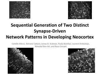
Sequential Generation of Two Distinct Synapse-Driven Network Patterns in Developing Neocortex
Sequential Generation of Two Distinct Synapse-Driven Network Patterns in Developing Neocortex. Camille Allene , Adriano Cattani , James B. Ackman , Paolo Bonifazi , Laurent Aniksztejn , Yehezkel Ben-Ari, and Rosa Cossart. Spontaneous Correlated Neuronal Activity.


Sequential Generation of Two Distinct Synapse-Driven Network Patterns in Developing Neocortex
E N D
Presentation Transcript
Sequential Generation of Two Distinct Synapse-DrivenNetwork Patterns in Developing Neocortex CamilleAllene, Adriano Cattani, James B. Ackman, Paolo Bonifazi, LaurentAniksztejn, Yehezkel Ben-Ari, and Rosa Cossart
Spontaneous Correlated Neuronal Activity • Developing cortical networks generate a variety of coherent activity patterns that participate in circuit refinement • Role in definition/construction of growing neural networks • Clarifying the underlying mechanisms and the spatiotemporal interactions between these diverse network patterns is crucial toward understanding their ultimate function in the construction of cortical maps
Typesof network patterns: cENO Cortical Early Network Oscillations • Large-scale oscillatory calcium waves • Occur immediately after birth at low frequency and providing most of the coherent activity in the developing rodent neocortex • Require action potentials • Driven by NMDA and AMPA but not GABAA receptors albeit GABA is a major excitatory neurotransmitter in the cortex at such early stages
Typesof network patterns: GDP Giant Depolarizing Potentials • Earliest synapse-driven network pattern in the developing hippocampus • Occur a few days after birth in rodents at moderate frequency (0.1 Hz). • Driven by GABAergic transmission • Disappear with the excitatory/inhibitory shift in the actions of GABA
Hypothesys • cENOs are cortical counterpart to the hippocampal GDPs but generated by glutamatergic synapses → GDPs and cENOs reflect intrinsic differences between brain structures OR: • cENOs and GDPs are separate network patterns sequentially dominating the developing neocortex
Methods • Functionalmultineuroncalcium imaging withtwo-photon laser scanning microscopy(average fluorescence within the cell vs time) → Characterizationof the events: rise times, amplitudes, and decay time constants • Electrophysiology: single-cell and field potential recordings targetedpatch-clamprecordings (whole-cellconfiguration) • Pharmacology. Antagonistsfor GABAA and ionotropicglutamatereceptors
Parameters to quantify synchronous activity patterns • Frequency: averaged time interval between two peaks of synchronous activity • Incidence: fraction of slices in which the pattern could be recorded at least once • Amplitude: average of the maximum of cells coactive in each peak of synchrony • Duration of synchronicity: number of successive frames for which the number of coactive cells was superior to threshold (reshuffling)
Maturation of population coherence in neocortical slices Maturation steps of spontaneous neuronal activity • E20: sporadic calcium spikes poorly correlated → immature action potentials • P0-P3: • cSPAs (Synchronous calcium plateaus) similar to hSPAs (not synapse driven: not affected by blocking AMPA/KARs, NMDARs, and GABAARs; blocked by sodium and L-type calcium channel antagonists) • cENOs associated with slow kinetics calcium events • P6-P8: cGDP confined within deeper cortical layers and always associated to fast calcium events
Multibeam two-photon imaging of the four maturation steps of spontaneous neuronal activity in somatosensory cortical slices from embryonic stages to first postnatal days Two-photon calcium fluorescence images of rat somatosensory cortical slices of the four types of spontaneous activity recorded at E20, P0 (cortical plate), P3 (cortical plate, horizontal slice), and P7 (deeper layers), respectively. scale bar: 100mm
Multibeam two-photon imaging of the four maturation steps of spontaneous neuronal activity Raster plots and fluorescent traces of the activity Black: calcium spikes; red: calcium plateaus (cSPA); green: cENO-events; blue: cGDP-events Current-clamp recordings (Vrest ~ 60 mV)
cENOs and GDPs display two distinct spatiotemporal dynamics Contour maps of 7 successive movie frames from a P3 and a P8 horizontalsomatosensory slice One frame every 150 ms; scale bar: 100m Slower dynamics of cENOs (1) compared with cGDPs (2) → large calcium waves vs fast calcium events
Fraction of imaged cells detected as being active for each movie frame in a P1 or P6 horizontal somatosensory cortical slice Calcium fluorescence traces of four cells implicated in the two cENOs illustrated in the above histogram Simultaneous field potential recording (FP) and calcium imaging (raster plot) Strong correlation between field potential oscillations and multineuron calcium activity Spectrogram of the FP oscillation associated to the cENO
cGDPvscENO • Peaks associated with cGDPs are • much smaller • more frequent • involve fewer cells • (raster plot) • than those associated with cENOs • cGDPare not associated with any remarkable oscillatory pattern but correspond to a significant increase in MultiUnitActivity as shown by the frequency histogram of MUA vs time and by the MUA recording trace
Single-cell electrophysiological and calcium events associated with cortical ENOs and GDPs Current-clamp recordings at resting membrane potential and corresponding calcium fluorescence traces of cells implicated in cENOs and cGDPs cENOs: calcium waves are associated with slowly rising and prolonged membrane potential depolarizations cGDP: calcium oscillations correspond to recurrent suprathreshold membrane potential depolarizations
Single-cell electrophysiological and calcium events associated with cortical ENOs and GDPs Comparison of three representative normalized calcium fluorescence traces recorded in single cells during cGDPs, cENOs, and cSPAs clearly illustrates the kinetics difference between these events. Fraction of calcium spike-, cSPA-, cENO-, and cGDP-cells relative to the number of active cells at four successive age groups between embryonic to first postnatal stages.
GABAergic transmission is not involved in the generation of cENOs but is crucial for cGDPs Fraction of imaged cells active for each movie frame as a function of time in a P3 and a P8 somatosensory horizontal slice Calcium fluorescence traces of 3 representative cells implicated in cENOs and cGDPs in control and after adding bicuculline cENOs are not affected by GABAAR blockade but completely prevented by AMPA/KAR and NMDAR antagonists
Differential role of glutamate in the generation of cortical ENOs and GDPs Fraction of imaged cells active for each movie frame vs time in a P0 somatosensory horizontal slice Occurrence of cENOs reduced/blocked with NMDAR/both NMDAR and AMPA/KAR antagonist Average i/V relationship of cENO-PSCs: Negative slope at hyperpolarized Em; Einv ~ 0 mV (NMDARs) Perfusion with the enzymatic glutamate scavenger GPT significantly reduces the frequency of cENOs; the effect of GPT is reversible upon wash out of the drug
Differential role of glutamate in the generation of cortical ENOs and GDPs Fraction of imaged cells active for each movie frame vs time in a P6 somatosensory horizontal slice Smaller effect on the occurrence of cGDPs compared with cENOs Blockade of ionotropic glutamatergic transmission almost completely prevented the occurrence of cGDPs → cGDPs also required glutamatergic transmission Linear i/V relationship of cGDP-PSCs; Einv ~ - 40 mV (GABAARs) • cGDPs frequency not affected by GTP but it decreased cGDPs amplitude (modified the fraction of cells Involved) • GPT does not affect cGDPs • as much as cENOs
Perfusion rate and anoxic/aglycemic episodes differentially affect cENOs and cGDPs P2 P7 P3 • The frequency of cGDPs was dramatically decreased • in low perfusion conditions • after 5 min of anoxia/aglycemia • The frequency of cENOs significantly increased • decreasing the rate of ACSF perfusion • after 5 min of anoxia/aglycemia Strong dependence of cENOs on glutamate levels NB: increases of extracellular glutamate concentration leading to transmitter diffusion are frequently associated to anoxic brain episodes
Differential modulation of cENOs and cGDPssimultaneously recorded in a neocortical slice. P4-P5 (Transition Period) Two types of synchronous network events: cGDPs → smaller amplitude highly recurrent synchronizations → fast and small amplitude calcium transients cENOs → less frequent large peaks of synchrony → slower and larger calcium transients Perfusion with anoxic/aglycemic ACSF increases the frequency of cENOs but reduces that of cGDPs cENOs and cGDPs are two distinct network patterns and not the expression of the same network pattern supported by different cellular mechanisms Representative calcium fluorescence traces from four imaged cells illustrating the amplitude and kinetics difference between cENO and cGDP-associated calcium events Average: amplitude difference between the two network patterns. Scaled average: significantly slower rise and decay time constants of cENOs-associated calcium transients than those associated to cGDPs
Differential modulation of cENOs and cGDPs simultaneously recorded in a neocortical slice. P4-P5 (Transition Period) • Comparison of control and perfusion with the enzymatic glutamate scavenger GPT (Alteration of the spatiotemporal glutamate profile without interfering with transmitter release or with glutamate receptors uptake mechanisms) • Perfusion with GPT selectively blocks the occurrence of cENOs (green) without significantly affecting cGDPs (blue) • → The action of glutamate during cENOs involve transmitter diffusion/accumulation in the extracellular space
CONCLUSIONS: cENO vs cGDP • ENOs and GDPs are two distinct network patterns, sequentially expressed in immature neocortical structures • Developmental profile: the expression of cENOs peaks around birth (P0 –P3) and they are no longer present when cGDPs dominate the network (P6 –P8) • Dynamics: • cENOs are low-frequency oscillations (0.01 Hz) displaying slow kinetics and gradually involving the entire network • cGDPs are recurrent oscillations (0.1 Hz) that repetitively synchronize localized neuronal assemblies • Mechanisms:cENOs are supported by NMDAR but not GABAAR activation, in contrast to cGDPs, and depend on extracellular glutamate concentrations
CONCLUSIONS: cENO vs cGDP • These two patterns are characterized by different spatiotemporal dynamics both in electrical and optical recordings • cENOs are effectively modulated by extracellular glutamate levels as • Short anoxic conditions facilitate cENOs • They are blocked by an enzymatic glutamate scavenger. • cENOs and cGDPs are two separate aspects of neocortical network maturation that may be differentially engaged in physiological and pathological processes
In vivo correlates • In vivo counterpart of cENOs is likely to be an endogenous brain rhythm expressed during sleep-like resting states and disappearing during the animal movement • Studies in neonatal rodents in vivo have characterized an early pattern of synchronized cortical electrical activity, the “spindle-bursts” that could be the in vivo expression of slice cGDPs as they share comparable dynamics, similarly confined spatial distribution and developmental profile
CURRENT CLAMP: VOLTAGE CLAMP: usefultofindinversionpotential → typeofchannel